Abstract
Background: Non-Alcoholic Fatty Liver Disease is the most increasing chronic liver condition with a global prevalence of 30%. Metabolic factors like Obesity, Diabetes Mellitus are considered as major risk factors. Complications include cirrhosis and hepatocellular carcinoma. The correlation between metabolic risk factors and NAFLD is known but needs to be highlighted through contextual evidence.
Objective: To investigate the frequency of various metabolic factors and their association with Alanine Transaminase levels in patients with Non-Alcoholic Fatty Liver Disease.
Method: It was a cross-sectional study carried out over 6 months in the Liver Center and Central Diagnostic Laboratory, Mayo Hospital, Lahore in 2023-24. A sample of 124 patients ultrasonically confirmed (Moderate to severe steatosis) was recruited and LFTs, lipid profile and fasting blood sugar were measured. Data was analyzed by SPSS v26. The descriptive statistics calculated, and Pearson correlation and simple linear regression were applied.
P value of <0.05 was considered significant.
Results: Out of 124 patients, 42 (34%) were males and 82 (66%) were females. The prevalence of NAFLD with raised ALT was 61.3% and with normal ALT was 38.7%. Central obesity in NAFLD patients was 98% according to waist circumference and 57.3% according to their BMI. 45% of NAFLD patients showed the features of Metabolic Syndrome while the remaining 55% did not. Elevated ALT levels were strongly correlated with the metabolic risk factors present in NAFLD patients (p = 0.019), repeated treatment (p = 0.049) as well as treatment failure (p < 0.001).
Conclusion: Investigation of various metabolic factors in diagnosed NAFLD patients suggests that Obesity is the most common metabolic factor followed by Type 2 Diabetes and then Hypertriglyceridemia. NAFLD showed a strong correlation to raised ALT levels, present in female population more than the male. Moreover, raised ALT levels in NAFLD patients also showed a strong association with metabolic risk factors.
Keywords: Non-Alcoholic Fatty Liver Disease, Metabolic Syndrome, Hypertension, Type 2 Diabetes
Introduction
Non-Alcoholic Fatty Liver Disease (NAFLD), also termed metabolic (dysfunction) associated fatty liver disease (MAFLD) is liver damage due to excessive fat accumulation in liver or hepatic steatosis in the absence of significant alcohol consumption or any other etiology like viral hepatitis [1]. It is diagnosed using either an imaging or a liver biopsy evaluation in suspected people with at least 5% hepatocyte infiltration with steatosis [2]. Classification of NAFLD includes two subtypes; nonalcoholic fatty liver (NAFL), the non-progressive form and NASH i.e. the progressive form of disease. NAFL is defined by histological evidence of hepatic steatosis with no features of hepatocyte injury including ballooning degeneration while, for the diagnosis of NASH, the presence of macro vesicular steatosis, inflammation, and ballooning hepatocytes is considered the histological criteria [3]. NAFLD is the most prevalent chronic liver problem around the globe as it affects 30% of the general global population while its prevalence in Asian countries is approximately 29.6% [4]. Lately, it is emerging as a global epidemic with increased risk of chronic liver diseases i.e. cirrhosis, steatosis (NASH) and most importantly hepatocellular carcinoma (HCC) [5]. NAFLD is believed to be the hepatic manifestation of metabolic syndrome [2]. The increased global incidence of NAFLD correlates with the rising
rates of MetS (Metabolic Syndrome) factors worldwide, particularly Obesity, Dyslipidemia and Type 2 Diabetes Mellitus (T2DM) resulting in the subsequently increased rates of morbidity and mortality due to metabolic, cardiovascular and hepatic diseases [6]. Obesity and Insulin Resistance are key trigger factors for both NAFLD and T2DM [7].
To diagnose NAFLD, liver biopsy is the gold standard but it cannot be widely used due to its invasive nature. As a result, noninvasive approaches i.e. imaging techniques such as abdominal ultrasound, CT, and MR and serum biomarkers particularly Alanine Transaminase levels (ALT) are widely used for diagnosis of NAFLD in recent years [8]. Serum Alanine Transaminase level depends directly on the incidence of metabolic syndrome. In NAFLD diagnosis, elevated serum ALT levels play a crucial role in indicating the presence of Obesity, Diabetes Mellitus, Hypercholesterolemia, Hypertriglyceridemia and low HDL levels. Each of these conditions independently affect hepatic ALT levels thus making it an excellent NAFLD screening marker [9]. Elevated ALT levels, with or without elevated AST (Aspartate Aminotransferase) can be used for NAFLD screening diagnosis. Even though some NAFLD patients may also present with normal ALT, thus decreasing its sensitivity for NAFLD detection. For this purpose, it is used along with Ultrasonography (US) for diagnosis of NAFLD [10]. If left undiagnosed, NAFLD can lead to cirrhosis eventually leading to carcinoma.
Usually, patients with normal ALT are left undiagnosed which can be very dangerous for such individuals. Moreover, even patients with elevated ALT levels don’t take NAFLD as a serious threat due to lack of awareness regarding NAFLD and its asymptomatic nature but it can prove to be lethal to their liver in the long run. Keeping in mind the aforementioned risks of NAFLD as a worldwide liver epidemic, this research aims to study the prevalence of metabolic factors contributing to NAFLD and their effect on ALT levels among the Pakistani community. To investigate the frequency of various metabolic factors and their association with Alanine Transaminase levels in Non-Alcoholic Fatty Liver Disease.
Method
This cross-sectional study was carried out in the Liver Center of Mayo Hospital in Lahore (MHL). Samples from 124 patients were collected, including both males and females between the ages of 18 – 70 years with abdominal ultrasound reports confirming the presence of fatty liver. Information was collected from these patients regarding demographics, clinical history, biochemical tests and their lifestyle. Waist circumference of the patients was measured using inches tape, blood pressure was measured using a sphygmomanometer, and height and weight were measured for calculating BMI. Data about lifestyle factors like physical activity and smoking was collected. Previous history of any surgery and hepatitis B and hepatitis C or any other known disease were recorded. All of the above information was collected with the help of a questionnaire attached. Patients diagnosed with fatty liver in ultrasonography (US) reports were subjected to blood routine chemistry tests primarily liver function tests (only ALT). The patients who presented with elevated or normal ALT levels were further screened for hepatitis B and hepatitis C. Patients who screened positive for hepatitis B or hepatitis C were excluded, while patients with negative screening underwent further routine chemistry blood analysis. The tests involved were blood sugar levels (fasting or random) and lipid profile (serum cholesterol levels, serum triglycerides levels, HDL-c levels).
The results of these parameters were analyzed as: ALT level = 0-40 U/L (Normal range) and values greater than 40 U/L were considered high. Cholesterol level = 0-200 mg/dL (Normal range) and values greater than 200 mg/dL were considered high. Triglycerides level = 0-150 mg/dL (Normal range) and values greater than 150 mg/dL were considered high. H.D.L-c = More than 40 mg/dL (males) and more than 50 mg/dl (females) (Normal range) and values less than 40 mg/dL (males) and less than 50 mg/dl (females) were considered low. Blood Sugar Fasting= More than or equal to 100 mg/dL were considered elevated. All of these routine chemistry blood evaluation tests were performed on Beckman Coulter AU-680 Chemistry Analyzer. Then, all the reports regarding measured parameters were analyzed. The frequencies of all these parameters i.e. Obesity, Waist Circumference, Age, Gender, BMI, Diabetes Type 2, Cholesterol levels, Triglycerides levels, HDL-c levels and systolic and diastolic blood pressures were calculated using SPSS and Correlation and Regression Analysis were carried out for different subgroups. Data was collected over six months after obtaining ethical approval from IRB (Institutional Review Board) (No.527/IRB/RC/KEMU) dated 17-11-2023.
Results
The study was conducted on 124 patients diagnosed with NAFLD in the Pakistani community. Out of these 124 patients, 42 (34%) were males and 82 (66%) were females. Among these patients, the mean
age was 46.84 years (SD = ±11.81). The highest frequency of the age range of patients diagnosed with NAFLD was 55-60 years among both sexes while the age range of 45-50 years and 35-40 years were the second most frequently present. The mean waist circumference was 102.24 (SD = ± 8.995) in males while in females it was
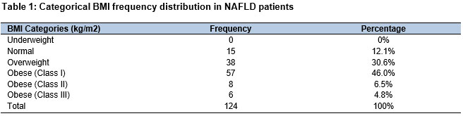
105.85 (SD = ± 14.149). Among 42 male subjects, 96% (40) males were represented with central obesity while 4% (2) males had no central obesity. Among a total of 82 female subjects included in our research, 80 (98%) had central obesity leaving only 2 females (2.4%) without central obesity (according to the NCEP ATP III criteria modified for Asians). In both sexes, 97% of NAFLD patients presented with central obesity with respect to waist circumference. The analysis of various metabolic factors showed that the most frequently reported BMI range was from 28.0-32.0 kg/m2. We categorized the NAFLD patients according to their BMI to demonstrate the relationship between the BMI and NAFLD in table 1. The table shows that there were more reported cases of NAFLD with higher BMI than in patients with relatively lesser BMI. Out of 124, no underweight NAFLD subject was reported. 15 (12.1%) had normal BMI. 38 (30.6%) were overweight, 57 (46.0%) were obese (class Ⅰ), 8 (6.5%) were Class Ⅱ obese and 6 (4.8%) were Class Ⅲ obese. The most reported cases of NAFLD were patients with class Ⅰ obesity out of which 42 were females and 15 were males. In total, 57.3% of obese patients presented with NAFLD.
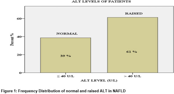
The evaluation of ALT levels among NAFLD patients suggested that the prevalence of NAFLD with raised ALT levels is more than normal ALT levels in the Pakistani population. Out of 124 subjects, only 48 (38.7%) had normal ALT levels while 76 (61.3%) had elevated ALT values. The mean ALT value among NAFLD patients was 44.66 (SD = ± 17.17).
Data gathered about fasting glucose levels (FPG) in NAFLD patients showed that 58 (46.8%) patients had raised FPG (fasting plasma glucose) levels and were diabetic out of which 17 (40.5%) were males and 41(50%)
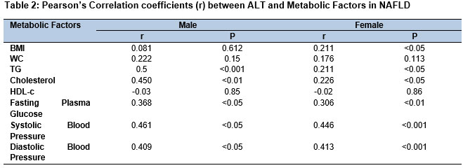
were females. The systolic and diastolic blood pressure analysis of patients showed that 47(37.9%) out of 124 were hypertensive. Among hypertensive patients included in our study, 13 (31%) were males and 34 (41.5%) were females. Lipid profile analysis of patients with NAFLD showed that 29 (23.4%) patients had elevated cholesterol levels including 8 (19%) males and 21 (25.6%) females. Hypertriglyceridemia was present in 54 (43.5%) NAFLD patients out of which 17 (40.5%) were males and 37 (41.5%) were females. Cut off values of HDL are different for males and females according to which 22 (52.4%) males and 27 (32.9%) females had lower than normal HDL-cholesterol levels. We compared the mean values of metabolic factors in male NAFLD patients with female NAFLD patients. Of the 124 patients with NAFLD enrolled in our study, using Pearson correlation formula, we found that there was a statistically significant correlation between the ALT values and most metabolic factors in each sex i.e. (p<0.05). The correlation between BMI and ALT levels in males was insignificant while in females it was significantly correlated. Additionally, all of the metabolic factors including TG (Triglycerides), FPG (Fasting plasma glucose), SBP (Systolic blood pressure), DBP (diastolic blood pressure) and cholesterol levels also had a significant positive correlation with the ALT levels in both sexes. Only WC (waist circumference) and ALT showed a weakly significant correlation as shown in Table 2 taking P value < 0.05 as significant.
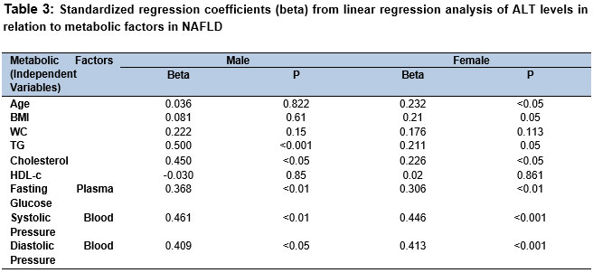
Moreover, Linear Regression Analysis was performed considering the ALT levels as the dependent variable and the values of all metabolic factors as the independent variable in each gender. The results are mentioned in the table below. The TG, FPG, SBP, DBP and cholesterol were independently associated with the ALT levels in both genders with significant P value <0.05. BMI was independently related to ALT levels in females of the Pakistani population while in males it lost its significance considering P value <0.05 as significant.
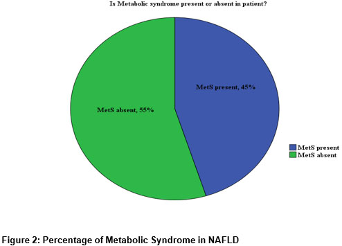
The frequency of metabolic syndrome in NAFLD patients was 45.2%. The frequency of patients having Metabolic Syndrome was 56 (55%) while 68 (55%) patients had no Metabolic Syndrome as shown in the figure below
Discussion
As can be seen from the results of this study, Non-Alcoholic Fatty Liver Disease or NAFLD has a high prevalence among the Pakistani community. It is positively correlated with central obesity and raised BMI. Moreover, ALT levels are also raised in NAFLD patients and show a strong correlation with the aforementioned metabolic factors [11–13]. To define the metabolic factors of MetS, we used the National Cholesterol Education Program Adult (NCEP) Treatment Panel III criteria modified for Asians. According to the criteria, MS is defined as the presence of at least three metabolic factors among the following: Abdominal obesity ≥90 cm (≥35.4 inches) for males and ≥80 cm (≥31.4 inches) for females or BMI >30 kg/m2, increased triglycerides levels (≥150 mg/dl), reduced HDL cholesterol levels (< 40 mg/dl) in men and (< 50 mg/dL) in women, increased blood pressure (≥ 130/85 mmHg), and Fasting Glucose (≥ 110 mg/dL or ongoing antihyperglycemic therapy) [14, 15]. According to this criteria, our research suggests that in Pakistani community, 57.3% of NAFLD patients had obesity with respect to their BMI, while according to waist circumference, 79.8% (99 patients) had central obesity, 47% (58 patients) were Diabetic, 37.9% (47 patients)
had high blood pressure, 43.5% (54 patients) had hypertriglyceridemia, 23.4% (29) had hypercholesterolemia and 39% (49 patients) had reduced HDL-cholesterol levels. The prevalence of MetS or metabolic syndrome was 45% in NAFLD patients which had 3 or more metabolic risk factors; while it was 55% in NAFLD patients who had only one or two metabolic risk factors present. NAFLD is the most frequent global cause of chronic liver disease, with a prevalence that differs from a global prevalence of around 30% in Western areas, 31·8% in the Middle East, 29.6% in Asian countries and 29.2% in the Chinese population [11, 12]. NAFLD is most likely driven by metabolic factors, dietary changes, physical activity, distribution of body fat and genetic makeup. NAFLD is the hepatic manifestation of MetS and thus has a bidirectional link with the factors of MetS including Obesity, Diabetes Type 2, Dyslipidemia and Hypertension. Previous studies show that NAFLD is diagnosed in 47–63 % of people with type 2 diabetes and about 80% of people with obesity, 76.6% of people with hyperlipidemia [13, 14]. In comparison to the worldwide trends of metabolic risk factors in NAFLD, we did research to find the prevalence of each metabolic factor in NAFLD patients. In patients with various chronic liver disorders, ALT levels usually reflect hepatic inflammation and liver injury. Most of the earlier studies suggest that the elevated ALT values were strongly correlated with the higher risk of NAFLD. However, in some other recent studies, NAFLD patients diagnosed by histology, MRI and ultrasonography possessed the normal ALT value. Normal ALT values hinder the timely diagnosis and treatment of NAFLD leading to advanced stage
i.e. NASH or liver fibrosis [16]. Thus, in our research, we calculated the prevalence of NAFLD patients possessing elevated and normal ALT levels in the Pakistani community. Our results showed that 61% of NAFLD patients had elevated ALT levels, while 39% of them had normal ALT levels. Previous studies show that the raised ALT levels in NAFLD patients are strongly correlated to metabolic factors and are usually lower in patients with no metabolic components present. They suggested that central obesity, raised FPG levels, TG levels and reduced HDL-c levels were strongly associated with raised ALT levels. Our research focused on observing the trends of this correlation between ALT levels and metabolic factors in the Pakistani population. We found a statistically strong correlation (p< 0.05) and (p<0.001) between most of these metabolic factors and ALT levels in NAFLD patients, except the BMI values of male subjects. HDL-c levels in females were also not strongly correlated with ALT values in the Pakistani population [17]. Moreover, there is significant evidence to suggest the importance of sex hormones in the occurrence of NAFLD. In females, Estrogen levels are proven to be protective, while raised testosterone levels increase the risk of developing NAFLD. On a global scale, research strongly suggests that the prevalence of NAFLD in males is much higher than in females [18]. However, research about NAFLD in Pakistan and neighbouring countries like India shows a higher prevalence of NAFLD in female patients [11]. In our research, the same trend can be seen. The prevalence of NAFLD was higher in females than in males. The possible explanation is that the mean age of females involved in this study was 48.13 ± 11.77. Most of the females included were menopausal. Moreover, in Pakistan, most of the females have a sedentary lifestyle and dietary habits of heavy fat intake due to which the chances of getting obese and developing NAFLD increases even before menopause. When we observed the physical activity of the subjects included in our research, we found that the highest frequency of people developing NAFLD had very little physical activity (less than an hour).
The study has limitations as it was a single-centre study, and NAFLD was screened through ultrasonography only. Another point was that the time convenience was followed, so this limited the sample size. This study is timely and has policy and practice implications.
Conclusion
Investigation of various metabolic factors in diagnosed NAFLD patients of the Pakistani community suggests that Obesity is the most prevalent metabolic factor followed by Type 2 Diabetes and then Hypertriglyceridemia. Other metabolic factors like Hypertension, low HDL-c levels and Hypercholesterolemia are prevalent with lesser frequencies. The prevalence of NAFLD showed a strong correlation to raised ALT levels, affecting the female population more than the male. Moreover, raised ALT levels in NAFLD patients also showed a strong association with metabolic risk factors.
References
- Navarroza AMC, Wong SN. Comparison of clinical and metabolic profiles of lean versus non-lean nonalcoholic fatty liver disease. Indian Journal of Gastroenterology. 2021;40(4):380-8. https://doi.org/10.1007/s12664-021-01184-6
- Lonardo A, Leoni S, Alswat KA, Fouad Y. History of Nonalcoholic Fatty Liver Disease. International Journal of Molecular Sciences. 2020;21(16):5888. https://doi.org/10.3390/ijms21165888
- Chalasani N, Younossi Z, Lavine JE, Charlton M, Cusi K, Rinella M, et al. The diagnosis and management of nonalcoholic fatty liver disease: practice guidance from the American Association for the Study of Liver Hepatology. 2018;67(1):328-57. https://doi.org/10.1002/hep.29367
- Batool S, Morton Cuthrell K, Tzenios N, Shehryar Z. Hepatocellular Carcinoma in Non-alcoholic Fatty Liver Disease: Emerging Burden. International Research Journal of Oncology. 2022;6(4):93-104. https://journalirjo.com/index.php/IRJO/article/view/116
- Wong S-W, Chan W-K. Epidemiology of non-alcoholic fatty liver disease in Asia. Indian Journal of Gastroenterology. 2020;39(1):1-8. https://doi.org/10.1007/s12664-020-01018-x
- Thomas JA, Acharya S, Shukla S, Thomas JJ, Pratapa SK, Hulkoti. Non alcoholic fatty liver disease (NAFLD) in metabolic syndrome (MetS)-A case control study. Med Sci. 2020;24(103):1490-9. https://discoveryjournals.org/medicalscience/current_issue/v24/n103/A66.pdf
- Targher G, Corey KE, Byrne CD, Roden M. The complex link between NAFLD and type 2 diabetes mellitus — mechanisms and treatments. Nature Reviews Gastroenterology & Hepatology. 2021;18(9):599-612. https://doi.org/10.1038/s41575-021-00448-y
- Kage M, Aishima S, Kusano H, Yano H. Histopathological findings of nonalcoholic fatty liver disease and nonalcoholic Journal of Medical Ultrasonics. 2020;47:549-54. https://doi.org/10.1007/s10396-020-01061-3
- Valle-Martos R, Valle M, Martos R, Cañete R, Jiménez-Reina L, Cañete MD. Liver enzymes correlate with metabolic syndrome, inflammation, and endothelial dysfunction in prepubertal children with obesity. Frontiers in Pediatrics. 2021;9:629346. https://doi.org/10.3389/fped.2021.629346
- Zou Y, Zhong L, Hu C, Sheng G. Association between the alanine aminotransferase/aspartate aminotransferase ratio and new-onset non-alcoholic fatty liver disease in a nonobese Chinese population: a population-based longitudinal study. Lipids in Health and Disease. 2020;19(1):1-10. https://doi.org/10.1186/s12944-020-01419-z
- Waseem M, Saeed F, Khan R. Ultrasound Detected Non-Alcoholic Fatty Liver Disease Prevalence and its Risk Factors in Pakistani Population. Advancements in Life Sciences. 2023;9(4):607-11. https://www.als-journal.com/9433-22/
- Zhou J, Zhou F, Wang W, Zhang XJ, Ji YX, Zhang P, et al. Epidemiological features of NAFLD from 1999 to 2018 in China. Hepatology. 2020;71(5):1851-64. https://doi.org/10.1002/hep.31150
- Golabi P, Paik J, Hwang JP, Wang S, Lee HM, Younossi ZM. Prevalence and outcomes of non‐alcoholic fatty liver disease (NAFLD) among Asian American adults in the United States. Liver International. 2019;39(4):748-57. https://doi.org/10.1111/liv.14038
- Purnell JQ. Definitions, Classification, and Epidemiology of Obesity. MDText.com, Inc., South Dartmouth (MA); 2000. https://www.ncbi.nlm.nih.gov/books/NBK279167/
- Prasad DS, Kabir Z, Dash AK, Das BC. Prevalence and risk factors for metabolic syndrome in Asian Indians: A community study from urban Eastern India. J Cardiovasc Dis Res. 2012;3(3):204-11. https://doi.org/10.4103/0975-3583.98895
- Powell EE, Wong VW-S, Rinella M. Non-alcoholic fatty liver disease. The Lancet. 2021;397(10290):2212-24. https://doi.org/10.1016/s0140-6736(20)32511-3
- Ma X, Liu S, Zhang J, Dong M, Wang Y, Wang M, et al. Proportion of NAFLD patients with normal ALT value in overall NAFLD patients: a systematic review and meta-analysis. BMC Gastroenterology. 2020;20:1-8. https://doi.org/10.1186/s12876-020-01165-z
- Nagral A, Bangar M, Menezes S, Bhatia S, Butt N, Ghosh J, et al. Gender Differences in Nonalcoholic Fatty Liver Disease. Euroasian J Hepatogastroenterol. 2022;12(Suppl 1):S19-S25. https://doi.org/10.5005/jp-journals-10018-1370