Abstract
Background: Uterine diseases are typically classified as neoplastic or non-neoplastic. When untreatable, they necessitate hysterectomy, with radiological and pathological examinations ensuring accurate diagnosis and improved patient care.
Objective: To describe and compare the clinical and radiological characteristics of uterine specimens of hysterectomy patients with histopathological findings.
Method: This cross-sectional study was carried out at Rashid Latif Medical College and its affiliated Arif Memorial Trust Hospital, comprising 300 hysterectomy specimens collected over a two-year period. A non-probability consecutive sampling method was used. Following ethical approval, data was obtained from medical records and histopathology requisition forms using a structured proforma. Clinical, radiological, and histopathological parameters were analyzed with SPSS version 25. Categorical variables, including clinical presentation, radiological diagnosis, type of procedure, and histopathological findings, were summarized as frequencies and percentages, whereas continuous variables were expressed as mean, standard deviation, and range.
Results: A total of 300 hysterectomy specimens were analyzed. The most frequent clinical indications were abnormal uterine bleeding (30%) and heavy menstrual bleeding (30%). Fibroids (35%) and adenomyosis (16.3%) were the leading histopathological findings. On descriptive comparison, radiological diagnoses matched histopathological findings with the highest match seen in fibroids (90.5%) and uterovaginal prolapse (100%).
Conclusion: Most histopathological diagnoses of post-operative hysterectomy cases were aligned with the radiological diagnosis. However, histopathological examination continues to be the definitive standard for diagnosing and excluding malignancy and is required for all surgical specimens.
Keywords: Hysterectomy, Radiology, Histopathology, Uterine
Introduction
The uterus is a vital female reproductive organ, essential for conception and childbirth. It nurtures and safeguards the growing baby, creating the ideal conditions for a healthy pregnancy and a successful birth [1]. Other than the major role of pregnancy, the uterus also contributes to complex hormone-regulated physiological processes. Estrogen is responsible for the proliferation of uterine epithelial cells, that ultimately lead to tissue growth, whereas progesterone is held responsible for restricting excessive proliferation. There is always a balance between these two hormones that is considered crucial for maintaining normal endometrial function. In uterus estrogen acts as the primary growth-stimulating factor [2]. Progesterone has dual function; it not only inhibits endometrial proliferation but can also lead to the development of uterine fibroids and other pathological conditions of uterus. Other than that, the response of uterus to hormonal stimulation is also determined by signaling of paracrine system between stromal and epithelial cells. It emphasizes the multiple roles including normal uterine physiology and disease states [3].
Diseases of the uterus can present with multiple clinical features that often causes problems and women have to take advise from the medical specialists. Although there are many problems but the most prevalent are the abnormalities in menstrual cycle, irregular abnormal uterine bleeding, lower abdominal pelvic pain, etc. these problems greatly affect the lifestyle of any women. So, they must take advice from the specialist, so that proper management can be done to improve the quality of life [4].
Hysterectomy is the most performed gynecological operation in many hospital settings included in our part of world. It is done for a variety of uterine disorders e.g fibroids, abnormal uterine bleeding, adenomyosis, and malignancies. The incidence of this type of procedure shows considerable variation across different regions of the world. Each surgical approach has its own advantages and limitations. While abdominal hysterectomy provides superior surgical exposure and is preferred in certain cases, it is associated with greater operative trauma and a higher risk of complications. Other than that, there is also availability of laparoscopic techniques. It is also widely used in gynecology. It is considered better because less complications are reported in this technique and reduced complication rates [5].
As far as radiological investigations are concerned, Ultrasound is commonly done procedure in hospital settings. Usually in routine practice it is commonly advised radiological test. It evaluates the proper management plan of different diseases especially related to uterus and pelvis. It is safe, noninvasive and commonly available diagnostic tool [6]. We should keep in mind that ultrasound is not always helpful to reach conclusive diagnosis. That is why all the biopsy specimens are sent for histopathological evaluation. It is considered gold standard. In many cases, especially related to gynecology, we need to do further immunohistochemistry. With the help of Histopathological examination, we can reach a conclusive diagnosis. It not only diagnoses the case but can also tell the exact morphology and depth of the lesions [7]. The important feature of histopathological examination is the differentiation between benign and malignant conditions of uterus and its adnexa. It is an essential component for further management [8].
There is a need for local evidence on how the Pakistani females present with gynecological uterine pathology and the correlation of radiological findings with histopathological diagnosis. Current study aims to describe the histopathological characteristics of uterine specimens of hysterectomy patients with radiological findings.
Method
This cross-sectional analytical study was conducted retrospectively at a private institution, Rashid Latif Medical College, and its affiliated hospital, Arif Memorial Hospital, Lahore. It was carried out in the Department of Pathology over a two-year period from November 2022 to October 2024. The total sample size was 300 hysterectomies, selected based on all eligible specimens, depending on the feasibility of available records. Data were collected using a structured proforma from patient medical records and pathology requisition forms, which included details such as age, clinical and radiological findings, reasons for hysterectomy, type of procedure performed, and correlation of radiological, clinical, and histopathological findings.
Inclusion criteria included all surgically resected hysterectomy specimens, including total abdominal hysterectomy (with or without salpingo-oophorectomy) and vaginal hysterectomy. Small biopsies, uterine fibroid samples, endometrial curettage samples, and autolyzed samples were excluded from the study.
Upon receipt in the Pathology Department, specimens were labeled, numbered, and fixed in 10% buffered formalin for 24–48 hours. Following a thorough gross examination, several sections were excised from key areas of the specimens, then processed into paraffin blocks. Sections of 4 microns were cut using a microtome and routinely stained with Hematoxylin and Eosin [9], [10].
Histopathological examination was performed on each hysterectomy specimen, with systematic observations recorded for the cervix, endometrium, myometrium, ovaries, and fallopian tubes. Tissue sampling and evaluation were conducted according to standard histopathological procedures, following the College of American Pathologists (CAP) protocol to ensure representative and clinically relevant sections from all anatomical sites were examined. Normal physiological states of the endometrium (proliferative, secretory, and atrophic) and conditions like chronic cervicitis and various functional ovarian cysts (including inclusion, follicular, luteal, and paratubal cysts) were classified as histologically ‘unremarkable’.
A detailed microscopic examination was carried out on the stained slides, and the lesions were classified into the following categories: lesions of the uterine corpus (including both endometrium and myometrium), lesions of the cervix, and lesions of the ovaries. The histopathological findings were subsequently compared with ultrasound results and clinical diagnoses. Ethical approval for the study was obtained from the Institutional Review Board (Reference No. IRB/2022/064, dated October 26, 2022). Data were analyzed using SPSS version 25.
Results
A total of 300 hysterectomy specimen reports were analyzed during the two-year study period.
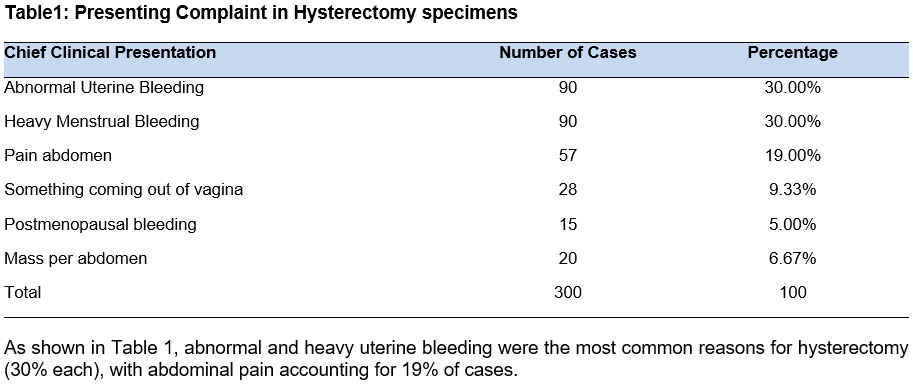
As shown in Table 1, abnormal and heavy uterine bleeding were the most common reasons for hysterectomy (30% each), with abdominal pain accounting for 19% of cases.
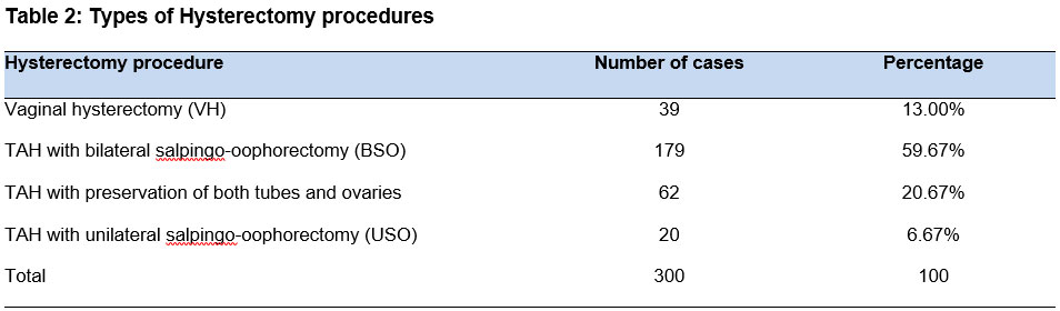
Table 2 illustrates that the most common procedure performed was abdominal hysterectomy with bilateral salpingo-oophrectomy, accounting for 179 cases (59.67%). Additionally, least common procedure was total abdominal hysterectomy with unilateral salpingo-oophrectomy was performed in 6.67% of cases.
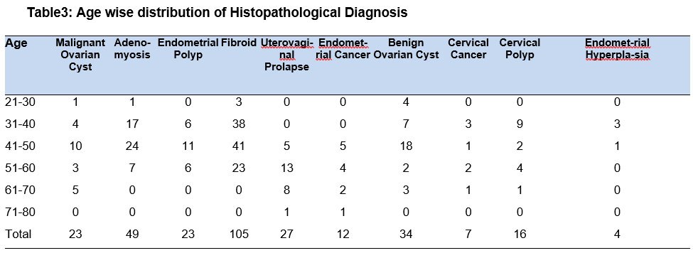
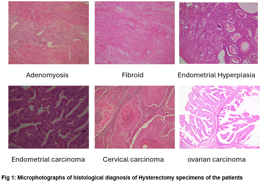
Table 3 indicates that fibroids were the most frequent pathology leading to hysterectomy, observed in 105 cases (35%), predominantly affecting the age group of 31 to 50 years. The least common pathologies resulting in hysterectomy were endometrial and cervical cancers, accounting for 3% and 2.3% of cases, respectively. Microphotographs are shown in figure 1
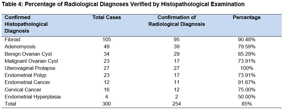
The above table shows that radiological findings corresponded with the results of histopathology in 85% of cases. The study compared ultrasound findings with histopathological diagnoses; other imaging modalities such as CT or MRI were not included. While clinical symptoms were recorded and analyzed descriptively, they were not part of the formal diagnostic correlation. Discrepancies between ultrasound and histopathology were noted in 15% of cases, particularly in conditions like adenomyosis and endometrial polyps.
Discussion
The study evaluated hysterectomy specimens to identify lesion patterns and aligned histopathological results with clinical indications and preoperative radiological diagnoses. Hysterectomy is a critical procedure in gynaecological practice for its effectiveness in resolving various health issues, thereby enhancing women's quality of life. Globally, it ranks as the second most frequently performed major surgical procedure among women, only surpassed by caesarean sections [11]. The rising rate of hysterectomies in developing countries is a growing concern and warrants closer evaluation. It is crucial to evaluate the clinical reasons and pathologies linked to hysterectomies performed on women [12].
In the present study, menstrual irregularities—specifically abnormal uterine bleeding and heavy menstrual bleeding—were the most common clinical indications for hysterectomy, accounting for a combined total of 60% of cases. It is reported in another study that abnormal uterine bleeding (AUB) is responsible for over 70% of gynaecological consultations among perimenopausal and postmenopausal women and is a major reason for them to seek medical help [13]. From this study, it is concluded that almost one third of hysterectomy cases revealed fibroids as the underlying cause. Histopathological examinations in the majority of studies consistently identify uterine leiomyoma as the commonest uterine tumor, observed worldwide as well as locally [14], [15].
In one study, leiomyoma was observed in 36.4% of women, adenomyosis in 21.8%, and endometrial hyperplasia in 3.6% [16]. These figures align closely with our findings, which identified fibroids in 35% of hysterectomy specimens, adenomyosis in 16.3%, and endometrial hyperplasia in only 1%. Endometrial and cervical cancers were among the least reported, consistent with findings from other studies [17].
Ultrasound is a commonly utilized diagnostic technique in gynaecology, known for its non-invasive and cost-efficient approach to evaluating different pathologies. However, the precision of ultrasound diagnostics can vary based on the specific pathology being examined, with histopathology frequently used as the definitive standard for verification [18]. In the present study, fibroids were correctly diagnosed in 90.48% of cases and adenomyosis in 79.59%, while another study demonstrated that ultrasound shows high diagnostic accuracy for identifying uterine fibroids and adenomyosis, with success rates of 96.25% and 98.75%, respectively [19]. Not only for benign conditions, ultrasound is also helpful in diagnosing gynaecological malignancies leading to hysterectomy; however, final diagnosis can only be established through histopathology [20].
In this study, we performed histopathological examination of the uterus and its adnexa using light microscopy in the laboratory. Ultrasound findings of these cases were obtained from clinical requisition forms. We compared histopathology results with ultrasound reports for all hysterectomy cases. In many cases, radiological and pathological findings matched, but there were several instances where diagnoses differed. Therefore, every hysterectomy specimen should be evaluated histopathologically to reach a conclusive diagnosis for proper management.
This study has certain limitations. As it is a single-center retrospective analysis, the findings may not be generalizable to other settings or populations. Secondly, the study relied solely on ultrasound as the radiological modality, which may have limited sensitivity in detecting certain pathologies such as adenomyosis or small lesions. Future studies should be multi-center and prospective, incorporating additional imaging modalities such as CT and MRI for stronger radiological correlation.
Conclusion
Most hysterectomies were performed for symptomatic benign conditions that were untreatable, with fewer cases conducted for malignancies, with overlapping clinical features. Most of the cases showed consistency between histopathological and radiological findings. However, given the inconsistency observed in several cases, the agreement between histopathological examination and preoperative radiological diagnosis becomes crucial. Consequently, histopathological analysis remains the definitive gold standard and should be mandatory for all surgical specimens to ensure accurate diagnosis and appropriate management.
References
- Kelleher AM, Demayo FJ, Spencer TE. Uterine Glands: Developmental Biology and Functional Roles in Pregnancy. Endocr Rev. 2019;40(5):1424–45. https://doi.org/10.1210/er.2018-00281
- Gupta AK, Gupta I, Suri AK. Histopathological Spectrum of Hysterectomy Specimens. J Adv Med Med Res. 2020;32(6):96–104. https://doi.org/10.9734/jammr/2020/v32i630440
- Marquardt RM, Kim TH, Shin JH, Jeong JW. Progesterone and estrogen signaling in the endometrium: What goes wrong in endometriosis? Int J Mol Sci. 2019;20(15). https://doi.org/10.3390/ijms20153822
- Kancharla DV, Sudhir DS. Adolescence and their recondite gynaecological issues. Int J Clin Obstet Gynaecol. 2022;6(6):96–8. https://doi.org/10.33545/gynae.2022.v6.i6b.1236
- Spaich S, Weiss C, Berlit S, Hornemann A, Sütterlin M. The hysterectomy: influence of the surgical method in benign disease on convalescence and quality of life. Arch Gynecol Obstet. 2023;307(3):797–806. https://doi.org/10.1007/s00404-022-06778-9
- Arezzo F, Cormio G, La Forgia D, Kawosha AA, Mongelli M, Putino C, et al. The Application of Sonovaginography for Implementing Ultrasound Assessment of Endometriosis and Other Gynaecological Diseases. Diagnostics. 2022;12(4). https://doi.org/10.3390/diagnostics12040820
- Ge Y, Yang S, Yu Y, Liu Y, Li R. Gynecopathology in the Diagnosis and Assessment of Chronic Endometritis. Clin Exp Obstet Gynecol. 2023;50(5). https://doi.org/10.31083/j.ceog5005093
- Scepanovic B, Andjelic N, Mladenovic-Segedi L, Kozic D, Vuleta D, Molnar U, et al. Diagnostic value of the apparent diffusion coefficient in differentiating malignant from benign endometrial lesions. Front Oncol. 2023;13(April):1–12. https://doi.org/10.3389/fonc.2023.1109495
- Li S. Chapter 22. Ben Cao Gang Mu. 2019;1897:684–741.
- Al-Taee RAM, Al-Aameli MH, Al-Qazwini YM. Histological Techniques: A brief Historical Overview. J Glob Sci Res. 2019;2(January):218–23. Available from: http://www.gsj-pub.comwww.gsj-pub.com/jgsr
- Soomro SB, Pirzada H, Zafar AF, Majeed M, Habib S. Indications and Histopathological Analysis of Hysterectomy Specimens. Pakistan J Med Heal Sci. 2022;16(7):319–20. https://doi.org/10.53350/pjmhs22167319
- Shahid R, Abbas H, Mumtaz S, Perveen F, Bari MF, Raja T, et al. Hysterectomy and Oophorectomy in Reproductive Age: A Cross-Sectional Study from a Tertiary Care Hospital. Cureus. 2020;12(5). https://doi.org/10.7759/cureus.8344
- Shrestha D, Aryal S, Tiwari A, Sharma R. Abnormal Uterine Bleeding among Women Visiting Gynecology Out-patient Department of a Tertiary Care Hospital: A Descriptive Cross-sectional Study. J Nepal Med Assoc. 2022;60(246):121–5. https://doi.org/10.31729/jnma.6954
- Zehra T, Gardezi SAB, Shams M, Zareen S, Khurshid IK, Irshad Z. Frequency and Spectrum of Non-Malignant Lesions in Abdominal Hysterectomy Specimens. J Bahria Univ Med Dent Coll. 2021;11(03):116–20. https://doi.org/10.51985/ZZYV1850
- Anshu, Gupta P, Singh T. The Concept of Self-Directed Learning: Implications for Practice in the Undergraduate Curriculum. Indian Pediatr. 2022;59(4):331–8. https://doi.org/10.1007/s13312-022-2501-x
- Rawat N, Tignath A, Jain A, Soni S, Agrawal M. Histopathological spectrum of uterine lesions in hysterectomy specimens. Int J Health Sci (Qassim). 2022;6(May):11176–80. https://doi.org/10.53730/ijhs.v6nS6.13085
- Mishra DA, Mishra DP, Kumar DBN, Pandey DT, Srivastava DS, Dwivedi DM. Histopathological spectrum of hysterectomy specimens and its correlation with clinical diagnosis at a tertiary care centre. Trop J Pathol Microbiol. 2019;5(4):240–7. https://doi.org/10.17511/jopm.2019.i04.10
- Grigore M, Popovici R, Himiniuc L, Scripcariu I, Toma B, Grigore A, et al. The added value of three‑dimensional ultrasonography in uterine pathology. Exp Ther Med. 2021;22(5):1–6. https://doi.org/10.3892/etm.2021.10696
- Shah SJH, Murrium SK-T-S, Naveed F, Fatima M, Gilani SA, Babar R. Comparative Study of Diagnostic Accuracy of Ultrasonography Vs Histopathology in patients Undergoing Hysterectomy due to Uterine Pathologies. Pakistan J Med Heal Sci. 2022;16(3):184–7. http://dx.doi.org/10.53350/pjmhs22163184
- Kumari A, Pankaj S, Choudhary V, Kumari J, Nazneen S, Kumari A, et al. Ultrasonic and Histopathological Evaluation to Exclude Premalignant and Malignant Lesions in Perimenopausal and Postmenopausal Women Presenting as Abnormal Uterine Bleeding. J Obstet Gynecol India. 2019;69(s2):171–6. https://doi.org/10.1007/s13224-018-1166-9