Abstract
Background: Rhegmatogenous retinal detachment (RRD) is a vision-threatening condition that requires timely surgical intervention to prevent permanent visual loss. Despite substantial advances in modern vitreoretinal surgery, the presence of proliferative vitreoretinopathy (PVR) continues to complicate management, often limiting anatomical success and functional recovery. Often, these cases need a drainage retinotomy to ease the tension and help reattach the retina. Conventional laser photocoagulation remains the standard method for sealing retinal breaks, but can induce thermal damage and postoperative scarring, limiting functional recovery. The use of human amniotic membrane graft (AMG) has recently attracted attention due to its anti-inflammatory, anti-fibrotic and regenerative properties that promote retinal healing and reduce fibrosis. International evidence supports the potential benefits of AMG in complex retinal detachment repair; However, local data is limited. This study was designed to compare the anatomic and visual outcomes of drainage retinotomy treated with laser alone versus laser combined with AMG in patients with RRD complicated by PVR. Objective: To report the anatomical and visual outcomes in RRD patients worsened by PVR, undergoing Retinotomy with LASER only vs patients undergoing Retinotomy with Amniotic Membrane Grafts and LASER.
Method: A prospective cohort study was conducted at the department of Ophthalmology, Lahore General Hospital over a period of twelve months from 1st feburary2023 to 1st February 2024. A total of 20 patients, 14 males (70%) and 6 females (30%) were included in this study. The patients were diagnosed case of rhegmatogenous retinal detachment complicated by PVR. We divided the patients into two groups; A and B. Group A underwent retinectomy with LASER only and Group B underwent Retinectomy with Amniotic Membrane Grafts and LASER. The patients were followed up for 3 months after surgery.
Results: Surgical outcome was measured in terms of achieving complete anatomical closure and improvement of visual acuity. In our study 100% of the patients achieved complete anatomical closure whereas visual acuity improvement was seen more in the patients treated with AMG than those treated with laser alone.
Conclusion: Visual and anatomical outcomes were better in patients that underwent AMG with LASER for the treatment of retinotomy as compared to those who underwent LASER only.
Keywords: RRD (rhegmatogenous retinal detachment), Retinotomy (Amniotic Membrane Graft), PVR (proliferative Vitreoretinopathy) LASER.
Introduction
Rhegmatogenous retinal detachment (RRD) is a dangerous surgical condition that, if left untreated, can cause major ocular morbidity. Retinal detachment is a sight threatening condition with an incidence of approximately 1 in 10000[1]. Prior to the 1920s, the condition was a permanent cause of blindness. Later, in Lausanne, Switzerland, Jules Gonin, MD, performed the first retinal detachment repair[2]. Repairing retinal detachments has been much easier and produced superior visual results in the past fifty years thanks to advancements in scleral buckling, pneumatic retinopexy, and vitrectomy procedures. The most significant peripheral retinal degeneration process that increases the risk of rhegmatogenous retinal detachment is thought to be lattice degeneration [3]. Through a number of methods, the retinal pigment epithelium (RPE) may normally keep its adherence to the neurosensory retina on top of it. These mechanisms include the interdigitation of the RPE microvilli and the outer segments of the photoreceptor, the metabolic activity of RPE, and the active transfer of subretinal fluid across RPE. When retinal detachment occurs, the neurosensory retina separates from the retinal pigment epithelium layer as a result of these processes becoming overloaded. When retinal detachment occurs, the neurosensory retina separates from the retinal pigment epithelium layer as a result of these processes becoming overloaded. Rhegmatogenous retinal detachment is the term for this condition. Retinal tears brought on by trauma or posterior vitreous separation are frequently the cause of rhegmatogenous retinal detachments. The distinctive look of rhegmatogenous retinal detachment sets it apart from tractional or serous detachments. Rhegmatogenous retinal detachment is diagnosed on clinical examination. Ocular coherence tomography and/or wide field fundus photography may be used to assess and record macula status. Additionally, B-scan ultrasound is recommended and could be a vital diagnostic tool in cases of media opacities. For rhegmatogenous detachments, surgical treatment is recommended. Identification, treatment, and closure of all retinal breaks are necessary for rhegmatogenous detachments. Repair options include vitrectomy, scleral buckle, laser retinopexy, pneumatic retinopexy, or a combination of these procedures. Despite advancements in modern vitreoretinal surgery, complex forms of retinal detachment (RD) with advanced proliferative vitreoretinopathy (PVR) in the context of chronicity or recurrence, traumatic RDs with retinal incarceration, and RD in proliferative vasculopathies continue to present difficult surgical scenarios [4]. Retinotomies and retinectomies help address traction and retinal shortening that continue after scleral buckling and membrane dissection. Retinectomy means "excising" the retina, but retinotomy means "cutting" or "incising" the retina. Retinectomy and retinotomy indications have grown to encompass a variety of difficult situations, providing a remedy in situations that initially appeared to be incurable. Since severe cryotherapy may raise the likelihood of epiretinal membrane formation by triggering retinal pigment epithelium cell release, laser treatment is usually used to cure the retinal break or breaks and prevent cyrotherapy, especially if many sites are required [5]. For many years, ocular surface diseases have been effectively treated with human amniotic membrane (hAM)[6]. hAM provides the best biological support for injured tissues and encourages recellularization because of its superior anti-inflammatory, anti-fibrotic, and antiangiogenic qualities as well as its low immunogenicity[7,8]. Research teams headed by Rizzo et al.[9] have reported successful in vivo applications of hAM for a variety of retinal diseases [10,11], including age-related macular neovascularization, posterior retinal fractures, retinal detachment (RD) [12] linked to MH, and refractory macular hole (MH) [13]. According to what is currently known, the application of hAM in vitreoretinal surgery is a new and creative technique with a lot of promise that requires more research and comprehension. Although AMG has been widely studied internationally, there is limited published data from Pakistan regarding its role in vitreoretinal surgery, which highlights the need for local evidence such as the present study.
Method
A prospective cohort study was conducted in the Department of Ophthalmology, Lahore General Hospital, from February 1, 2023, to February 1, 2024. The study enrolled 20 consecutive patients diagnosed with rhegmatogenous retinal detachment (RRD) complicated by proliferative vitreoretinopathy (PVR). The sample size was calculated using the WHO sample size calculator, assuming a 95% confidence interval, 80% power, and an expected difference of 30% in visual outcome between the two groups based on previous literature.
Inclusion Criteria: Patients of either sex, aged 18–70 years, with a confirmed diagnosis of rhegmatogenous retinal detachment complicated by proliferative vitreoretinopathy at any stage, and who provided written informed consent, were eligible for inclusion.
Exclusion Criteria: Patients with a previous history of retinal surgery in the affected eye, traumatic retinal detachment, active ocular infection, or systemic conditions known to impair wound healing such as uncontrolled diabetes mellitus or ongoing immunosuppressive therapy were excluded.
Sampling Technique and Group Allocation: A non-probability consecutive sampling technique was employed. Twenty eligible patients were enrolled and equally allocated into two groups (n=10 each). Group A underwent drainage retinotomy with LASER photocoagulation alone, while Group B received drainage retinotomy combined with an amniotic membrane graft (AMG) in addition to LASER treatment.
Data Collection: Baseline data included demographic characteristics, preoperative fundus examination findings, and B-scan ultrasonography. Postoperative assessments were conducted at one week, one month, and three months to evaluate anatomical retinal reattachment and changes in visual acuity.
Statistical Analysis: Data were entered and analyzed using SPSS version 25. Quantitative variables, including improvement in visual acuity, were expressed as mean ± standard deviation and compared between groups using the independent samples t-test. Categorical variables, such as anatomical closure rates, were presented as frequencies and percentages and analyzed using the Chi-square test. A p-value of <0.05 was considered statistically significant.
The Ethical Board Approval was taken from AMC, Lahore General Hospital under the reference number 102/LGH/2023 dated 06-01-2023.
Results
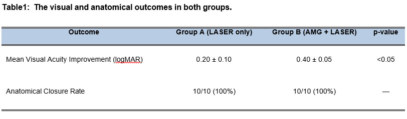
A total of 20 patients (14 males, 6 females; mean age 47 ± 8 years) were included and divided into two equal groups (n=10 each). Group A underwent drainage retinotomy with LASER only, while Group B underwent drainage retinotomy with amniotic membrane graft (AMG) and LASER.
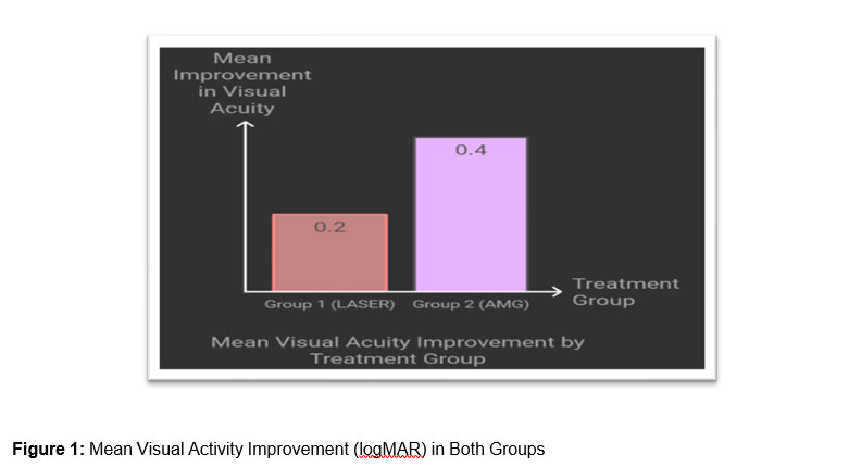
Visual Outcomes: Group A showed a mean improvement in visual acuity of 0.20 ± 0.10 logMAR, whereas Group B showed a greater mean improvement of 0.40 ± 0.05 logMAR. The difference between groups was statistically significant (p < 0.05).
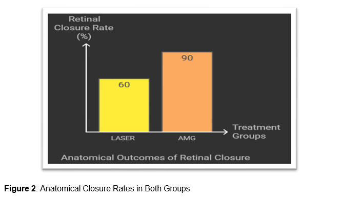
Anatomical Outcomes: All patients in both groups (100%) achieved complete anatomical closure at 3-month follow-up.
Discussion
Retinal detachments, diabetic retinopathy, and macular holes are among the retinal disorders that can be treated surgically by retinotomy. In order to improve retinal reattachment or healing, an incision is made into the retina to release mechanical strain. Amniotic membrane grafts and laser photocoagulation therapy are the two primary methods used to treat retinotomy. Although both approaches have been extensively researched, the decision between them is based on the patient's results, the underlying condition, and the surgeon's preference. The effectiveness, safety, and results of laser-treated retinotomy are contrasted with those of amniotic membrane grafts in this discussion.
An established treatment for a number of retinal conditions is laser photocoagulation [14]. In a study conducted by Lesley A Everett Retinal laser photocoagulation is still an essential therapeutic approach for the treatment of diabetic retinopathy and diabetic macular edema, even with the extensive usage and great effectiveness of anti-VEGF therapy [15].
This method can help seal retinal tears, lessen vascular leakage, and stop more retinal detachment by burning the retina with a laser. Retinal vein occlusions, diabetic retinopathy, and early-stage retinal detachment are among the disorders for which it is frequently employed. The less intrusive aspect of laser photocoagulation is one of its main benefits. The operation takes minimal time to recuperate from, is done in an outpatient setting, and is rather straightforward. By producing adhesions between the retina and underlying tissue through a controlled heat effect, it lowers the chance of separation and stops fluid from building up behind the retina. Although laser photocoagulation is effective in selected retinal conditions, its utility is limited in more complex clinical scenarios. Extensive retinal detachments, large retinal breaks, or macular involvement may respond suboptimally to laser treatment alone. Surgical outcomes are also influenced by operator expertise and laser parameters, as inadequate application can result in complications such as retinal scarring, macular injury, or suboptimal visual recovery. In advanced cases, including large retinal tears or longstanding detachment, additional surgical interventions may be required to achieve stable reattachment. Furthermore, laser-treated eyes, particularly those with extensive patholog, remain at risk of developing proliferative vitreoretinopathy, characterized by fibrocellular proliferation and recurrent traction on the retina.
Amniotic membrane grafting has emerged as a useful adjunct in the management of complex retinal pathologies where conventional approaches may be insufficient [16]. Chen et al. reported a markedly lower rate of recurrent retinal detachment in eyes treated with human amniotic membrane, with all graft-treated eyes remaining anatomically stable, compared with a high re-detachment rate in the control group that required additional surgical intervention [17]. The amniotic membrane, derived from the innermost placental layer, contains collagen-rich extracellular matrix components, growth factors, and anti-inflammatory mediators that support tissue repair and modulate fibrosis. Owing to these properties, its application has expanded across ophthalmic surgery, including the management of diabetic retinopathy, macular holes, retinal detachment, and corneal surface disorders. When used as an intraocular graft, the membrane provides a biological scaffold that promotes retinal healing, facilitates closure of retinal defects, and may reduce the risk of recurrent separation compared with laser-based techniques alone [15]. These regenerative characteristics are particularly advantageous in cases involving macular pathology or complex detachment associated with advanced retinal disease.
One of the principal advantages of amniotic membrane transplantation lies in its capacity to support retinal tissue regeneration [18]. In contrast to laser therapy, which may induce thermal injury and subsequent scarring, amniotic membrane grafts facilitate tissue repair with a reduced fibrotic response and more physiological restoration of retinal architecture [19]. This difference in healing response may translate into improved functional outcomes, particularly in eyes with extensive retinal breaks or chronic macular holes that have failed to respond adequately to laser treatment. Evidence from clinical studies indicates that the use of human amniotic membrane is associated with superior anatomical stability and a lower incidence of recurrent detachment when compared with laser-based approaches alone [20]. Additionally, when incorporated as an adjunct to procedures such as pars plana vitrectomy, amniotic membrane grafts appear to create a favorable microenvironment for retinal repair by limiting inflammation and promoting cellular recovery.
Despite its therapeutic advantages, amniotic membrane grafting is not without limitations. Although the procedure has demonstrated a favorable safety profile, it is technically more demanding and requires surgical expertise in the handling and placement of biological grafts. Accurate positioning of the membrane is essential to achieve stable retinal adhesion and optimal outcomes. While the risk of complications such as infection or graft-related inflammatory reactions is low, these possibilities must be considered, particularly in complex cases. In addition, the clinical applicability of this technique may be constrained by limited availability of processed human amniotic membranes in certain settings.
While amniotic membrane grafting offers clear therapeutic benefits, certain practical limitations should be acknowledged. The procedure, although generally safe, is technically demanding and requires specific surgical experience in the preparation and intraoperative placement of biological graft material. Precise positioning of the membrane is critical, as suboptimal placement may compromise retinal adhesion and surgical success. Although complications such as infection or graft-related inflammatory responses are uncommon, they remain relevant considerations, particularly in eyes with advanced or complex pathology. Furthermore, the broader implementation of this technique may be limited in some clinical settings due to restricted access to adequately processed human amniotic membranes.
Both laser photocoagulation and amniotic membrane grafting demonstrate clinical utility in the management of retinotomy, with their roles largely determined by disease complexity and intraoperative findings. Laser-based treatment remains an effective and dependable approach for uncomplicated retinal tears and early detachments. In contrast, amniotic membrane grafting appears to offer a valuable alternative in more complex retinal pathology, particularly in eyes with extensive retinal breaks or macular involvement where conventional laser therapy alone may be insufficient. The type of retinal pathology, the surgeon's background, and the particular requirements of the patient all influence which of these two approaches is best. It is probable that these therapy modalities will continue to be improved as retinal surgery develops, with the possibility of merging the two methods to enhance patient outcomes.
Our findings are consistent with previously published international studies; however, there is a paucity of local literature on the use of amniotic membrane grafts for drainage retinotomy in Pakistani patients [17]. A study conducted by Khan et al. (Pakistan Journal of Ophthalmology, 2022) reported improved anatomical success rates when AMG was combined with vitrectomy in complex retinal detachments. Our results strengthen these observations by demonstrating superior visual outcomes with AMG even in a smaller prospective cohort.
This study demonstrates several methodological strengths, including its prospective design, the use of objective outcome measures such as logMAR visual acuity and anatomical closure rates, and a standardized postoperative follow-up protocol. Nevertheless, certain limitations should be acknowledged. The relatively small sample size, the single-center nature of the study, and the short duration of follow-up may limit the generalizability of the findings. Extended follow-up and larger, multicenter studies would be valuable to assess the long-term stability of retinal reattachment and to identify delayed complications, including recurrence of proliferative vitreoretinopathy.
The findings of the present study indicate that the use of amniotic membrane grafts in combination with LASER during drainage retinotomy is associated with improved functional outcomes in patients with rhegmatogenous retinal detachment complicated by proliferative vitreoretinopathy. This approach could be considered particularly in cases where conventional LASER alone may not yield optimal visual recovery. Future studies with larger multicenter cohorts and extended follow-up are recommended to confirm these findings and to explore cost-effectiveness and patient-reported quality of life outcomes in our population.
Conclusion
Combining amniotic membrane graft (AMG) with LASER for drainage retinotomy resulted in superior visual outcomes compared to LASER alone, with 100% anatomical success in both groups. This combined approach may be considered a preferred option for managing complex rhegmatogenous retinal detachment cases complicated by proliferative vitreoretinopathy (PVR), although larger multicenter studies with longer follow-up are recommended to validate these findings.
Acknowledgements:
The author(s) acknowledge the use of AI-based tools for language editing and clarity enhancement. The final content, interpretation, and conclusions remain the sole responsibility of the author(s).
References
- Haimann MH, Burton TC, Brown CK. Epidemiology of retinal detachment. Arch Ophthalmol. 1982;100(2):289- https://doi.org/10.1001/archopht.1982.01030030291012
- Gonin J. The treatment of detached retina by searing the retinal tears. Arch Ophthalmol. 1930;4(5):621- https://doi.org/10.1001/archopht.1930.004500621
- Lewis H. Peripheral retinal degenerations and the risk of retinal detachment. Am J 2003;136(1):155-60. https://doi.org/10.1016/S0002-9394(03)00144-2
- Wykoff CC, Schwartz SG, Adelman RA, Brucker AJ, Flynn HW. Primary rhegmatogenous retinal detachment repair: evidence supports an individualised approach. Br J Ophthalmol. 2015;99(11):1451- https://doi.org/10.1136/bjophthalmol-2015-307651
- Warren A, Wang DW, Lim JI. Rhegmatogenous retinal detachment surgery: A review. Clin Exp Ophthalmol. 2023;51(3):271-9.
- De Rötth A. Plastic repair of conjunctival defects with fetal membranes. Arch Ophthalmol. 1940;23(3):522- https://doi.org/10.1001/archopht.1940.00860130586006
- Niknejad H, Yazdanpanah G, Kakavand M. Extract of fetal membrane would inhibit thrombosis and hemolysis. Med Hypotheses. 2015;85(2):197-202. https://doi.org/10.1016/j.mehy.2015.04.030
- Tseng SC, Espana EM, Kawakita T, et al. How does amniotic membrane work? Ocul Surf. 2004;2(3):177-87.
- Caporossi T, Pacini B, De Angelis L, Barca F, Peiretti E, Rizzo. Human amniotic membrane to close recurrent, high myopic macular holes in pathologic myopia with axial length ≥ 30 mm. Retina. 2020;40(10):1946-54.
- Caporossi T, Pacini B, Bacherini D, et al. Human amniotic membrane plug to promote failed macular hole closure. Sci Rep. 2020;10(1):18264. https://doi.org/10.1111/aos.14109
- Caporossi T, De Angelis L, Pacini B, Tartaro R, Finocchio L, Barca F, Rizzo A. Human Amniotic Membrane plug to manage high myopic macular hole associated with retinal detachment. Acta Ophthalmol. 2020;98(2):e252-6. https://doi.org/10.1111/aos.14174
- Caporossi T, Tartaro R, De Angelis L, Pacini B, Rizzo S. A human amniotic membrane plug to repair retinal detachment associated with large macular tear. Acta Ophthalmol. 2019;97:821-3. https://doi.org/10.1097/IAE.0000000000002699
- Caporossi T, De Angelis L, Pacini B, Rizzo S. Amniotic membrane for retinal detachment due to paravascular retinal breaks over patchy chorioretinal atrophy in pathologic myopia. Eur J Ophthalmol. 2020;30:392-5. https://doi.org/10.1177/1120672119891415
- Ramamurthy SR, Dave VP, Chou HD, et al. Retinotomies and retinectomies: A review of indications, techniques, results, and complications. Surv Ophthalmol. 2023;68(5):639-59. https://doi.org/10.1016/j.survophthal.2023.06.012
- Everett LA, Paulus YM. Laser therapy in the treatment of diabetic retinopathy and diabetic macular edema. Curr Diab Rep. 2021;21(9):1-2. https://doi.org/10.1007/s11892-021-01403-
- Iannetta D, Chhablani J, Valsecchi N, et al. Epiretinal implant of human amniotic membrane to treat highly myopic macular hole retinal detachments: A novel surgical technique. Eur J Ophthalmol. 2024;34(1):304-9. https://doi.org/10.1177/11206721231206717
- Khaqan HA, et al. Amniotic Membrane Graft for the Treatment of Giant Refractory Macular Hole Closure. Pak J Ophthalmol. 2022;38(3):212-7. https://doi.org/10.1097/IAE.0000000000003505
- Chen Y, Chen. Human amniotic membrane grafts for retinal breaks in diabetic tractional retinal detachment. Sci Rep. 2021;11(1). [Retracted] https://doi.org/10.1038/s41598-021-86804-z
- Ferreira MA, Maia A, Machado AJ, et al. Human amniotic membrane for the treatment of large and refractory macular holes: a retrospective, multicentric, interventional study. Int J Retina Vitreous. 2021;7(1):38. https://doi.org/10.1186/s40942-021-00308-6
- Zeydanlı EÖ, Özdek Ş, Yalçın E, et al. Human Amniotic Membrane: A Seal for Complex Retinal Detachments. Turk J Ophthalmol. 2024;54(5):268-74. https://doi.org/10.4274/tjo.galenos.2024.56424