Abstract
Background: Dengue is an arboviral infection caused by four dengue virus serotypes (DENV 1–4), with a global incidence that disproportionately affects tropical and subtropical regions. Despite a significant disease burden in Pakistan, local data on the hematological and biochemical profiles of different dengue categories is limited and not generating reliable evidence for clinical decision making.
Objective: To evaluate differences in clinical features, hematological indices, and biochemical parameters across classic dengue (CD), dengue hemorrhagic fever (DHF), and severe dengue (SD) and to corelate findings with severity of disease.
Method: A cross-sectional study design was conducted from Nov. 2021 to Nov. 2022, at three tertiary care military hospitals in Pakistan. 131 inpatients aged 14 years or older with confirmed dengue infection via NS1 antigen or serology, who presented within 72 hours of fever onset were eligible and included in the study. Systemic diseases and concurrent infections were excluded. Data were collected using a standardized tool capturing clinical features and laboratory parameters (hemoglobin, hematocrit, platelet count, total leukocyte count). Data were analyzed with SPSS v24.0, employing descriptive statistics and inferential tests (chi-square, one-way ANOVA) at a 95% confidence level.
Results: Of the 131 patients (62% male, 38% female), the highest frequency seen in the 15–30-year age group. Platelet count emerged as a significant marker of severity, with SD cases showing the lowest mean platelet counts (p<0.001). Leukopenia was observed in 84% of cases, though hemoglobin and hematocrit largely remained within normal limits across all dengue categories. DHF cases demonstrated the highest frequency of bleeding manifestations (10%). No mortality was recorded in the study population.
Conclusion: The study elucidated distinct hematological, clinical and biochemical profiles across dengue categories. The platelet count proved to be a reliable, low-cost marker for early severity grading, complemented by the consistent presence of leukopenia. These findings provide local data to refine triage and management protocols, addressing a significant gap in the diagnostic approach to dengue in Pakistan’s resource-constrained healthcare settings.
Keywords: Platelets, Hemoglobin, Hematocrit, Dengue hemorrhagic fever (DHF), Classic dengue (CD)
Introduction
Dengue is a type of arbovirus transmitted to humans via arthropod vectors, particularly the Aedes aegypti. It is caused by four genotypes of the dengue virus (DENV 1, DENV 2, DENV 3, DENV 4) [1,2]. This hyperendemic disease has a global occurrence of up to 390 million every year, out of which only 96 million exhibit dengue clinically, especially in tropical and subtropical urban or semi-urban areas of Asia and America, according to estimates from the World Health Organization (WHO). As of 2021, dengue continues to prevail in South America, India, Kenya, Peru, Philippines, Vietnam and some European countries [3,4].
All DENV serotypes may have the same mechanism of action that can lead to symptoms ranging from mild fever to lethal dengue shock syndromes. Hence, their virological characteristics have categorized the DENV infection into three main types known as dengue fever (DF), dengue hemorrhagic fever (DHF) and dengue shock syndrome (DSS) [5,6]. Dengue disease progression begins with a febrile phase where patients experience high fever, headaches, vomiting, flu and arthralgia for approximately 7 days. The next stage involves the critical phase which is a life-threatening contingency of acute symptoms such as internal bleeding and plasma leakage. However, the patient recovers when the vascular permeability is restored, but abnormal permeability could induce a sudden hypovolemic shock (DSS) if the critical phase is not controlled [6].
Although dengue may be distinguished via clinical symptoms, there are a variety of tests to confirm diagnosis, such as anti-DENV antibodies IgG, DENV-specific nucleic acid detection, and non-structural protein 1 (NS1) antigen test which is the most used. This will help provide affordable healthcare and treatment, especially to patients with atypical symptoms. Dengue-specific tests are usually not required for acute management cases but should still be done according to WHO [7,8].
Precise and early diagnosis is imperative for proper treatment to avoid complications of this disease. In developing countries, healthcare facilities may lack sophisticated diagnostic equipment; therefore, basic blood tests can still help in providing valuable clinical insights through important hematological and biochemical details. Patients suffering from dengue have been reported with a consistent altered biochemical profile that includes, but is not limited to: thrombocytopenia, leukopenia, elevated hematocrit, coagulopathy, and changes in lipid profile and liver enzymes. These parameters are helpful to make clinical decisions and can aid in estimation of the severity and classification of diseases such as dengue fever, dengue hemorrhagic fever and dengue shock syndrome [9–12].
Locally, patients suffering from dengue are not coming to tertiary healthcare, along with detailed biochemical and hematological examinations during the various stages of the disease. This lack of local data is detrimental to the ability to standardize the diagnostic protocols and triage tools used, tailored to the healthcare system in our region. In this study, we intended to fill the gap by conducting a descriptive cross-sectional study to assess the clinical, biochemical and hematological profiles of patients who were clinically diagnosed with dengue fever and admitted in tertiary hospitals. This study analyzes, evaluate, and interpret critical parameters using low-cost markers, considering the financial limitations of an underdeveloped healthcare framework. This can enable timely intervention and grade the severity level through early diagnosis.
Method
This cross-sectional study was conducted from 1st November 2021 to 30th November 2022 at three tertiary care military hospitals in Pakistan: Combined Military Hospital (CMH) Lahore, CMH Multan, and the Armed Forces Institute of Pathology (AFIP). All patients included were inpatients, and the patient flow during the study period was moderate. Inclusion criteria were patients aged 14 years or older, with no known systemic diseases, and normal baseline hematological and biochemical parameters prior to dengue infection. Exclusion criteria included the presence of systemic disorders (e.g., chronic kidney or liver disease, autoimmune disorders), bacterial or fungal infections, inflammatory diseases (e.g., tuberculosis, rheumatoid arthritis), diabetes mellitus, transplant patients, blood disorders (e.g., thalassemia, leukemia), malignancies, and COVID-19 infection. Secondary data of all eligible patients during the study period was included in the study.
The medical records of 131 patients diagnosed with dengue infection were reviewed, 60 from CMH Lahore, 40 from CMH Multan, and 31 from AFIP. Diagnosis was confirmed via positive NS1 antigen test, with patients presenting within 72 hours of fever onset and clinical features consistent with dengue. The study population included 81 males (62%) and 50 females (38%) aged between 14 and 74 years, primarily comprising military personnel and civilians from rural backgrounds. The sample size was determined based on the availability of eligible inpatient dengue cases during the study period. However, to ensure adequacy, the sample size was also estimated using Slovin’s formula, which yielded a minimum required sample size of 124 (assuming a population size of 600 and a margin of error of 8%). The final sample included 131 cases, exceeding the estimated requirement. Data was collected using a pre-designed data extraction tool that included clinical history, physical examination, laboratory reports, and monitoring sheets. Laboratory parameters assessed included hemoglobin, hematocrit, total leukocyte count, and platelet count. Vital signs such as temperature, blood pressure, and pulse were logged manually, and dengue-related symptoms like rash and bleeding were recorded using a structured clinical observation checklist.
A pilot test on 20 patients was conducted to ensure clarity and consistency of the tool. Data collection procedures were standardized across all centers and validated by infectious disease specialists, with trained medical officers and nurses responsible for consistent recording. All data were cross-verified with official medical records. Ethical approval was obtained on 29th February 2024 from the Institutional Review Board (IRB) of CMH Lahore Medical College & Institute of Dentistry (750-ERC/CMH/LMC dated 29/2/24). Patient confidentiality and data protection were maintained throughout the study. Statistical analysis was performed using SPSS version 24.0. Descriptive statistics, including frequencies and percentages, were used to summarize qualitative variables such as gender, age groups, and dengue classifications. For quantitative variables such as hemoglobin level, platelet count, total leukocyte count, and hematocrit percentage, the mean ± standard deviation (SD) was calculated. The association between gender and dengue types was assessed using the chi-square test. Normality of the numerical data was evaluated using the Spearman test. One-way ANOVA was applied to compare the means of quantitative variables across different dengue categories. All statistical analyses were conducted at a 95% confidence level, and a p-value of <0.05 was considered statistically significant. No missing data were encountered during the analysis.
Results
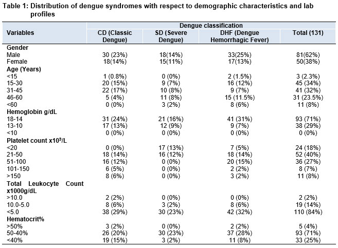
Out of 131 confirmed dengue cases, 62% were males and 38% were females. Dengue Hemorrhagic Fever (DHF) was more prevalent among males (n=33), whereas Classic Dengue (CD) was more common among females (n=18). The highest proportion of cases occurred in the 15–30-year age group (34%), followed by 31–45 years (32%). Severe Dengue (SD) was most frequent in the 46–60 age group. Refer to Table 1. Hemoglobin levels were largely preserved, with 71% of patients falling in the normal range (14–18 g/dL); no cases had Hb <10 g/dL. Most patients (84%) demonstrated leukopenia (<5 x10³/μL), and 71% had hematocrit levels within the normal range (40–50%). A quarter of the patients (25%) had hematocrit levels <40%. Thrombocytopenia was prominent, with 40% of patients showing platelet counts between 21–50x 109/L and 18% with counts below 20 x 109/L. Refer to Table 1 for details.
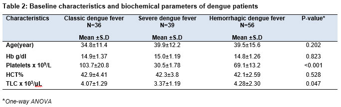
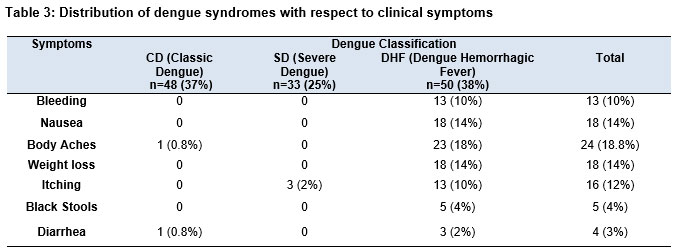
In Table 2, significant differences were found in platelet count (p<0.001) and total leukocyte count (TLC) (p=0.047) across dengue severity groups. Classic dengue had the highest platelet count, while severe dengue had the lowest. Hemorrhagic dengue had the highest TLC. Age, hemoglobin, and hematocrit showed no significant differences. DHF patients had the highest frequency of bleeding symptoms (10%), and other common clinical features included body aches (18.8%), nausea (14%), weight loss (14%), and itching (12%). Notably, 18.3% of patients who tested negative for the NS1 antigen were diagnosed with dengue based on clinical signs, thrombocytopenia, and IgM/IgG ELISA results. Refer to Table 3.
Discussion
At the end of our study, we found that no mortality occurred in our study population (n=131). It should be noted that the age group 16-30 years old showed the highest prevalence of dengue cases, which is supported by the fact that this trend increases with age [13]. Another aspect to this may include the need for outdoor exposure, especially for the male population where males were more affected than females, indicating that males are mostly breadwinners and involved in outdoor activities. This further reciprocates on the fact that the hospital setting is military-based, where the majority of faculty are male, and the respective patients admitted were mostly soldiers by occupation.
A gender-based study conducted in Western Uttaranchal Pradesh also reached the same conclusion of dengue prevalence to be more dominant in the male population, as the cultural backgrounds of Pakistan are quite like India [13]. However, in terms of prevalence of each dengue type, more females 15 out of 50 (30%) as compared to males 18 out of 81(22%) were infected with severe dengue which was also noted in two studies done in China [14] and Bangladesh [15] where majority of the patients were found to be 15-30 years old.
An accurate diagnosis of dengue must be made precisely. For instance, in patients with DHF, platelet and hematocrit levels are generally used to diagnose dengue severity, whereas leukocyte count helps to identify type of causation (viral or bacterial) [16]. In our study, 93 patients (71%) with DENV infection with or without warning signs had normal hemoglobin levels and normal hematocrit. These figures were like that of a study done at a hospital in Bangkok, Thailand [17]. Based on incidental findings, a significant relationship between Hemoglobin and HCT% was also proved at another medical institute in Thailand in March 2021 [18]; however, the resource for a three-fold conversion method to determine Pearson’s R2 with respect to RDW (RBC Distribution Width) is lacking.
A study in South Africa defined their patients with platelet count less than 50x109/L as having severe thrombocytopenia, whereas those patients above 50x109/L were termed to have non-severe thrombocytopenia. They observed in their study that a significantly lower number of patients had severe thrombocytopenia during the febrile phase, critical phase, and recovery phase [19]. However, our study showed quite the opposite results, where 58% of patients had platelet counts less than 50x109/L, whereas 42% of patients had platelet counts above 50x109/L/L. This difference could be possible due to environmental and climate changes. Leukopenia is a condition resulting from bone marrow suppression due to the production of cytokines secreted as opposed to a direct or indirect viral infection [16]. In our study, over 110 patients (84%) suffered from leukopenia but within normal limits. Out of the two patients in our study with total leukocyte count greater than 10,000 g/dL, no patient suffered from severe shock which occasionally is observed if leukocytosis occurs as was also observed in two studies in 2020 done by Triana and Dessy [16] and another study done by Wayez A and Zafar L [20].
Since our study is a cross-sectional analysis, our research has some limitations. Our sample represents only 3 hospitals and a specific population group so external validity might be somewhat compromised. Conducting study on a larger sample size accurately represents the diversity of our population. A larger sample of dengue patients can help analyze a wider range of demographics and geographic variables.
Conclusion
This study successfully characterized the clinical, hematological and biochemical profiles of dengue patients across different disease categories; Classic Dengue (CD), Dengue Hemorrhagic Fever (DHF), and Severe Dengue (SD), admitted to tertiary care hospitals. Our findings confirm that platelet count is a critical and cost-effective hematological marker, significantly decreasing with increasing disease severity (highest in CD, lowest in SD), thus serving as an important tool for early severity grading and clinical management. Leukopenia was common across all groups but was notably higher in DHF cases, reflecting immune suppression during disease progression. Hemoglobin and hematocrit values remained largely within normal limits and did not significantly differ among dengue classifications, suggesting limited utility for severity assessment in this context. Clinically, bleeding manifestations were more frequent in DHF patients, aligning with the biochemical profile and underlining the importance of integrated clinical and laboratory evaluation. The predominance of cases in the 15–45-year age group highlights the vulnerability of the working-age population, possibly due to environmental exposure and vector prevalence in these demographics. By delineating distinct hematological patterns corresponding to dengue categories, this study addresses the need for accessible, low-cost diagnostic markers to assist timely triage and management in resource-limited healthcare settings. These insights contribute valuable local data that can inform diagnostic protocols and improve patient outcomes in similar endemic regions.
References
- Harapan, H., Michie, A., Sasmono, R. T., & Imrie, A. (2020). Dengue: A minireview. Viruses, 12(8), 829. http://doi.org/10.3390/v12080829
- Chala, B., & Hamde, F. (2021). Emerging and re-emerging vector-borne infectious diseases and the challenges for control: A review. Frontiers in Public Health, 9. https://doi.org/10.3389/fpubh.2021.715759
- Kularatne, S. A., & Dalugama, C. (2022). Dengue infection: Global importance, immunopathology and management. Clinical Medicine (London, England), 22(1), 9–13. http://doi.org/10.7861/clinmed.2021-0678
- Dengue and severe dengue. (n.d.). Who.int. Retrieved March 21, 2024, from https://www.who.int/news-room/fact-sheets/detail/dengue-and-severe-denguehttps://www.who.int/news-room/fact-sheets/detail/dengue-and-severe-dengue
- Sunil, S., N. Sirisena, P. D. N., Mahilkar, S., Sharma, C., & Jain, J. (2021). Concurrent dengue infections: Epidemiology & clinical implications. The Indian Journal of Medical Research, 154(5), 669. https://doi.org/10.4103/ijmr.ijmr_1219_18
- Kok, B. H., Lim, H. T., Lim, C. P., Lai, N. S., Leow, C. Y., & Leow, C. H. (2023). Dengue virus infection – a review of pathogenesis, vaccines, diagnosis and therapy. Virus Research, 324(199018), 199018. http://doi.org/10.1016/j.virusres.2022.199018
- Raafat, N., Blacksell, S. D., & Maude, R. J. (2019). A review of dengue diagnostics and implications for surveillance and control. Transactions of the Royal Society of Tropical Medicine and Hygiene, 113(11), 653–660. https://doi.org/10.1093/trstmh/trz068
- Fongwen, N., Wilder-Smith, A., Gubler, D. J., Ooi, E. E., T. Salvana, E. M., de Lamballerie, X., Olliaro, P. L., & Peeling, R. W. (2021). Target product profile for a dengue pre-vaccination screening test. PLoS Neglected Tropical Diseases, 15(7), e0009557. http://doi.org/10.1371/journal.pntd.0009557
- Wang, W.-H., Urbina, A. N., Chang, M. R., Assavalapsakul, W., Lu, P.-L., Chen, Y.-H., & Wang, S.-F. (2020). Dengue hemorrhagic fever – A systemic literature review of current perspectives on pathogenesis, prevention and control. Wei Mian Yu Gan Ran Za Zhi [Journal of Microbiology, Immunology, and Infection], 53(6), 963–978. https://doi.org/10.1016/j.jmii.2020.03.007
- Mutanabbi, M., Shova, S. S., Kibtiar, M., & Mosleh, T. (2022). Clinical profile and lab findings of dengue fever in children admitted in a tertiary care hospital. Mymensingh Medical Journal: MMJ, 31(3), 741–748.https://pubmed.ncbi.nlm.nih.gov/35780359/
- Vijay, J., Anuradha, N., & Anbalagan, V. P. (2022). Clinical presentation and platelet profile of dengue fever: A retrospective study. Cureus. https://doi.org/10.7759/cureus.28626
- Ananda Rao, A., R, R., Gosavi, S., & Menon, S. (2020). Dengue fever: Prognostic insights from a complete blood count. Cureus. https://doi.org/10.7759/cureus.11594
- Macias, A. E., Werneck, G. L., Castro, R., Mascareñas, C., Coudeville, L., Morley, D., Recamier, V., Guergova-Kuras, M., Etcheto, A., Puentes-Rosas, E., Baurin, N., & Toh, M.-L. (2021). Mortality among hospitalized dengue patients with comorbidities in Mexico, Brazil, and Colombia. The American Journal of Tropical Medicine and Hygiene. https://doi.org/10.4269/ajtmh.20-1163
- Verma, R., Kumar, M., & Mishra, B. (2020). Prevalence of dengue fever in Western Uttar Pradesh, India: A gender-based study. InternSational Journal of Applied & Basic Medical Research, 10(1), 8. https://doi.org/10.4103/ijabmr.IJABMR_337_18
- Yang, J., Mosabbir, A. A., Raheem, E., Hu, W., & Hossain, M. S. (2023). Demographic characteristics, clinical symptoms, biochemical markers and probability of occurrence of severe dengue: A multicenter hospital-based study in Bangladesh. PLoS Neglected Tropical Diseases, 17(3), e0011161. https://doi.org/10.1371/journal.pntd.0011161
- Haider, N. (2023). Bangladesh’s 2023 Dengue outbreak-age/gender-related disparity in morbidity and mortality and geographic variability of epidemic burdens. International Journal of Infectious Diseases, 136, 1–4. https://doi.org/10.1016/j.ijid.2023.08.026
- Triana, D., Kurniati, A., & Wirastari, G. G. (2020). Relationship Between Platelet, Hematocrit and Leukocyte with Dengue Severity in (Vol. 7). https://www.researchgate.net/publication/348694543_European_Journal_of_Molecular_Clinical_Medicine
- Riswari, S. F., Budiman, M. F., Darmayanti, D., Ernawati, Prodjosoewojo, S., Susandi, E., Oehadian, A., & Alisjahbana, B. (2022). A comparison of the accuracy of handheld hemoglobinometer and hematocrit measurements for detecting plasma leakage in dengue hemorrhagic fever. International Journal of General Medicine, 15, 2589–2595. https://doi.org/10.2147/ijgm.s343017
- Malik, J. (2023). Association of pattern of thrombocytopenia and serology with timings of plasma leakage in patients of dengue hemorrhagic fever during dengue epidemic 2019-an experience from Rawalpindi Medical University: A cross sectional study. The Professional Medical Journal, 30, 461–466. https://doi.org/10.29309/TPMJ/2023.30.04.7362
- Yasuda, I., Saito, N., Suzuki, M., Umipig, D. V., Solante, R. M., Guzman, F. D., Sayo, A. R., Yasunami, M., Koizumi, N., Kitashoji, E., Sakashita, K., Ng, C. F. S., Smith, C., & Ariyoshi, K. (2021). Unique characteristics of new complete blood count parameters, the Immature Platelet Fraction and the Immature Platelet Fraction Count, in dengue patients. PloS One, 16(11), e0258936. http://doi.org/10.1371/journal.pone.0258936