Abstract
Background: Type 2 Diabetes Mellitus (T2DM) is a significant worldwide and national public health concern, with a high frequency observed in clinical settings. HbA1c is used as a normal for evaluating glycemic control but does not indicate the underlying chronic inflammation connected to T2DM pathophysiology. The NLR, a simple and low-cost inflammatory marker derived from a routine complete blood count, has been associated with T2DM and its complications in numerous populations, but its diagnostic utility in the Pakistani population remains inadequately explored.
Objective: To find the frequency of T2DM in patients presenting to the outpatient department (OPD) and to compare the mean Neutrophil-Lymphocyte Ratio (NLR) between patients with and without T2DM.
Method: This cross-sectional was conducted at King Edward Medical University in Lahore. The sample of 323 consecutive participants visiting the OPD was enlisted. Demographic and clinical information was made. The blood samples were tested on HbA1c and complete blood count. Participants were divided into T2DM (HbA1c 5.7 and above) and Non-Diabetic (HbA1c 5.69 and below). An independent samples t-test was used in the comparison of the means in NLR in the two groups. Gender and age stratification of data was also performed.
Results: T2DM patients were in high frequency in this sample i-e 49.8% (161/323). The average NLR of the T2DM group (2.51 ± 0.84) was also significantly increased over non-diabetic group (1.64 ± 0.54) at <0.001 p-value. This major disparity was maintained in cases where the data were stratified based on age and gender.
Conclusion: The neutrophil-lymphocyte Ratio in the T2DM patients is much higher than that in non-diabetic patients. Simple and cheap NLR is a marker with a close correlation to existence of T2DM.
Keywords: Neutrophil-Lymphocyte Ratio; Type 2 Diabetes Mellitus; Glycemic Control; HbA1c; Inflammation.
Introduction
Diabetes Mellitus (DM) is a major health problem in the world as its prevalence is steadily rising throughout the world. The estimates of the International Diabetes Federation show that there were 463 million individuals with diseases in the world in 2019, and in 2045, the figure will be 700 million [1]. This epidemic puts a heavy burden on the healthcare systems of all countries, including the developing nations including Pakistan. The national disease burden in Pakistan is very alarming, as recent statistics show that about 27.4 million people have the disease, and the situation is marked by a significant national crisis in the sphere of public health [2].
Type 2 Diabetes Mellitus (T2DM) is an increasing beta-cell dysfunction and insulin resistance disease that comprises the vast majority of patients with diabetes [3]. Microvascular and Macrovascular complications, such as coronary artery disease, neuropathy, retinopathy, and nephropathy are major causes of chronic hyperglycemia in T2DM [4]. These complications are the most frequent reasons for mortality and morbidity, and coronary artery disease is the deadliest one [5]. The fundamental means of prevention or postponement of these sequelae lies in the realization and optimal glycemic control [6].
Glycated hemoglobin (HbA1c) is the gold standard of evaluating long-term Glycemic control and is a measure of the average level of glucose in the blood over the past three months [7]. Nevertheless, HbA1c has its limitations; it fails to detect the changes in glucose and is not reliable during the condition of red blood cells turnover. More importantly, it does not capture the underlying, long-standing, low grade inflammatory condition which is currently being established as a core pathophysiological constituent of T2DM and complications of the condition [8].
The NLR is an inexpensive, simple, inflammatory biomarker that can be determined using a conventional complete blood count that is becoming a noteworthy indicator of systemic inflammation. This is a composite measure because it reveals the activity of both the adaptive and the innate immune pathway. Modern studies have attributed high NLR rates to poor glycemic control and an improved risk of developing diabetic complications [9]. In support of this, a systematic review found a significant difference, and the NLR was significantly lower in patients through poor glycemic control than those with good control [10]. The diagnostic precision of NLR to assess glycemic control in the Pakistani population, however, is a poorly studied subject. Thus, the present study is supported by the necessity of a convenient, cheap inflammatory biomarker that can be used to supplement HbA1c, especially in resource-constrained areas such as Pakistan. The determination of the diagnostic utility of NLR may give the clinical society an extra instrument to detect individuals with high risks and who might need more intensive management, potentially enhancing patient outcomes. This study was therefore designed with the following objectives:
- To find the frequency of T2DM in patients presenting to
- To compare mean NLR in cases with and without
Method
This study was designed as a cross-sectional study that was conducted collectively in the Endocrinology and Pathology departments of Mayo and King Edward Medical University Hospital in Lahore. It was a three-month research investigation, which began once the research synopsis had been formally approved. A total of 323 people were recruited to participate in the study. This sample size was determined to be the minimum requisite, calculated based on a 95% confidence interval, an absolute precision of 5%, and an anticipated population percentage (frequency) of Type 2 Diabetes Mellitus (T2DM) of 70%, as cited in a previous study [11]. All eligible patients who presented to the outpatient department throughout the study duration were invited to participate until the required sample size was reached using non-probability.
Inclusion and exclusion criteria were as follows. Inclusion criteria encompassed patients of both genders aged between 18 and 75 years who were presenting to the OPD for a routine checkup. Exclusion criteria were a diagnosis of type 1 diabetes; HbA1c levels in the pre-diabetic range (5.7-6.9%); the presence of acute infections, as evidenced by neutrophilic leukocytosis or clinical signs; diagnosed thyroid disorders; chronic liver or kidney disease; any rheumatological diseases; any known malignancy; current use of immunosuppressive therapy; and pregnancy.
After obtaining informed consent, clinical profile and demographic data, including name, body mass index (BMI), gender, age, and history of smoking, dyslipidemia, hypertension, and anemia, were recorded using a specially designed proforma. Recent HbA1c values were obtained from patient records or tested accordingly. Venous blood samples were drawn under aseptic measures using a 3cc disposable syringe. Samples were analyzed for HbA1c and a complete blood count using a standard hematology analyzer (Manufacturer: e.g., Sysmex Corporation, Kobe, Japan). The absolute neutrophil and lymphocyte counts were obtained from the automated differential leucocyte count. The absolute neutrophil count was considered divided by the absolute lymphocyte count to find the NLR.
All the data was statistically processed using SPSS Statistics (Version 26.0). The Shapiro-Wilk test was used to test the normality of the quantitative data. In the case of continuous variables, e.g., age, NLR and Body Mass Index (BMI), the outcomes are given as the mean values with standard deviation (SD). Categorical variables, such as gender and T2DM status are summarized as counts and per centages. The proportion of prevalence of T2DM, which is characterized by a level of 7 or above in HbA1c was computed relative to the total population in a study. To compare the mean NLR in the diabetic (HbA1c ≥7%) and non-diabetic (HbA1c <5.7%) groups, an independent samples t-test was applied. The data was also stratified based on age (age of 50 years and above versus age of 50 years and below) and gender to control possible confounders. After stratification, a t-test was done on the results of NLR among the T2DM and non-diabetic groups in each stratum. The ethical approval of this investigation was done by the King Edward Medical University Institutional Review Board/ Ethical Review Committee and CPSP with a p-value of 0.05. (RTMC# HEM-2022-066-944, Date of Approval: 01/08/2025). All participants were given patient consent before they could be enrolled into the study.
Results
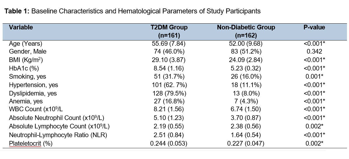
In this cross-section analysis, 323 participants were involved. The frequency of T2DM (HbA1c ≥7%) in our sample was 49.8% (161/323). Table I includes a summary of the baseline characteristics of the patients. The mean Body Mass Index (BMI) of the T2DM group was much higher (p=0.001). Hypertension, dyslipidemia, and anemia were found to be notably more common in the group of T2DM (p<0.001).
Comparison of Mean NLR
One of the main objectives of the research was to compare the difference among the mean of the NLR in the two cohorts. The statistical analysis showed that the mean NLR was significantly advanced in T2DM group as it was 2.51 (SD = 0.84), as compared to the non-diabetic population where it was 1.64 (SD = 0.54) (p<0.001).
Stratification Analysis
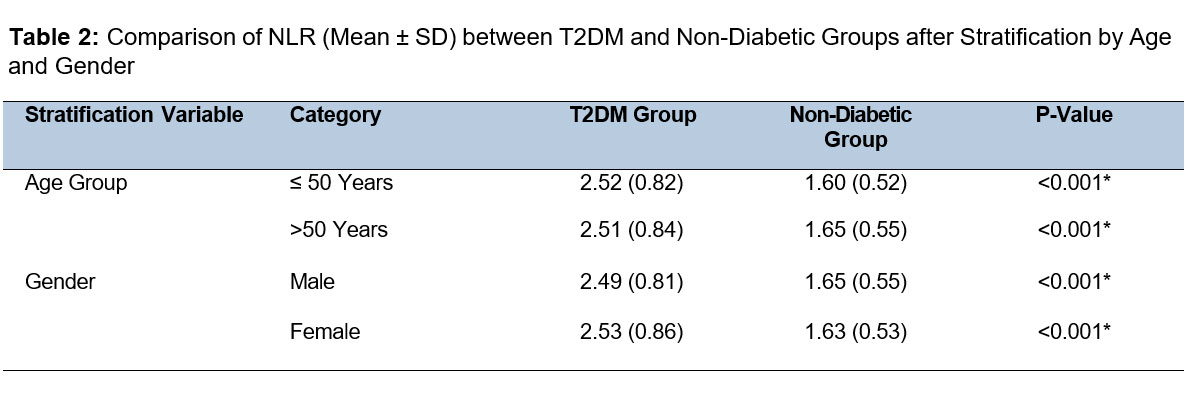
Stratification of data was done in terms of age and gender as intended. The mean NLR revealed that there was considerable rising in the T2DM group associated to the non-diabetic group among strata as revealed in Table II.
Discussion
The aim of this cross-sectional research was to find out the prevalence of T2DM and the evaluation of the NLR of diabetic and non-diabetic individuals presenting to a tertiary care OPD in Lahore, Pakistan. We found out that T2DM was frequent in our clinical sample (49.8%). What was more important was that we discovered statistically significant increase in the average NLR between the T2DM patients (2.51 ± 0.84) and non-diabetic (1.64 ± 0.54), which was consistent across the age and gender stratifications.
The critical increase in NLR in our T2DM group is in strong agreement with the growing amount of international data that has positioned systemic inflammation as being central to T2DM pathophysiology [10]. We are not alone in our findings since a complete cross-sectional analysis of the NHANES data showed that, despite a complete adjustment of various confounding factors, high levels of NLR were independently related to a high risk of T2DM, which supports the validity of our results with other populations [11,12]. The finding that this large disparity persisted following age and gender stratification in our study is further evidence of the strength of NLR as a strong inflammatory indicator and is not substantially diluted by age and gender.
The pathophysiological explanation of this correlation is well-grounded. Pro-inflammatory state with activation of innate immune system pathways is facilitated by chronic hyperglycemia [13]. The major agents of innate immunity, neutrophils, are activated and increase in number releasing proteases and reactive oxygen species that lead to endothelial dysfunction and insulin resistance [14]. At the same time, adaptive immunity is also dysregulated, which is often expressed in the form of relative lymphocytopenia, which may be the result of enhanced apoptosis caused by the effect of inflammatory cytokines and hyperglycemic stress. These two opposing immunological pathways are thus combined into one powerful indicator of the systemic load of inflammation by the NLR [16].
The findings that we obtained are obtained from a clinical perspective when combined with the evidence that has shown that NLR can be associated with glycemic control and diabetic complications. The meta-analysis and systematic review that particularly addressed this relationship discovered that the standardized mean difference of NLR between patients with good and poor glycemic control was 0.79, which demonstrated significantly higher NLR in patients with poorly managed T2DM [17]. The same analysis showed that poor glycemic control was related to increase in NLR (OR=1.70). This implies that the high NLR in our T2DM group is not just an indicator of clinical diabetes but perhaps even of the extent of glycemic derangement and underlying inflammatory pathology, which is not adequately estimated by HbA1c alone.
In addition to glycemic control, the prognostic usefulness of NLR is also applicable in the field of microvascular complications. An experiment conducted on diabetic nephropathy proved that the NLR values were significantly elevated in groups of norm-albuminuria, microalbuminuria, and macroalbuminuria [17]. Moreover, NLR demonstrated a good predictive quality of microalbuminuria, and the area under the curve was 0.869 [18]. This demonstrates that NLR might be used as a risk marker at an early stage at low cost to detect diabetic patients at risk of developing nephropathy. The utility of NLR is not limited to microvascular complications; the prognostic strength of NLR in the macrovascular disease is graphic. A cohort study of patients who received percutaneous coronary intervention identified that patients with T2DM and high NLR obtained the poorest clinical results, with a significantly higher rate of serious adverse cerebrovascular and cardiovascular events [19]. This synergistic effect underlines the utmost significance of inflammatory status determination with the use of NLR among diabetic patients to have a complete stratification of cardiovascular risks. The strengths of our research are as follows: the research is prospective, patient groups were well characterized, and their exclusion criteria were strict to reduce confounding issues, and the gold standard of diagnosing and categorizing T2DM is used based on the HbA1c. Our findings can be highly relevant to practice in a basic hospital setting, especially where inflammatory panels are costly, such as Pakistan, where we show that the NLR can be calculated, using a basic CBC, and at very low costs.
Nonetheless, this does not mean our study is free. First, it is single-center and cross-sectional, which excludes the possibility of causality and does not ensure the generalizability of the results to the population of Pakistan as a whole. And secondly, we failed to conduct oral glucose tolerance tests, which would have excluded those with isolated postprandial hyperglycemia. Third, the unavailability of diabetic complications and long-term outcomes do not allow correlating NLR with the progression of the disease or end-organ damage in our cohort. Further longitudinal and large-scale studies are required to obtain the clinical actionable cutoff values of NLR and establish a temporal relationship between NLR and the development of T2DM and its outcomes in the Pakistani population.
Conclusion
This paper has shown that there is a very high NLR among T2DM patients as compared to normal people. This is a non-complicated inflammatory marker, the NLR, which is based on a standard complete blood count and is highly related to T2DM. By virtue of its possible ability to indicate the state of glycemic control and the probability of developing diabetic comorbidities, the NLR ought to be viewed as an effective adjunctive biomarker in the overall treatment of T2DM.
References
- Williams R, Karuranga S, Malanda B, et al. Global and regional diabetes prevalence estimates for 2019 and projections for 2030 and 2045: Results from the International Diabetes Federation Diabetes Atlas. Diabetes Res Clin Pract. 2019;157:107843. https://doi.org/10.1016/j.diabres.2019.107843
- Basit A, Fawwad A, Qureshi H, Shera. Prevalence of diabetes, pre-diabetes and associated risk factors: second National Diabetes Survey of Pakistan (NDSP), 2016–2017. BMJ Open. 2018;8(8):e020961. https://doi.org/10.1136/bmjopen-2017-020961
- American Diabetes Association. Classification and diagnosis of diabetes: Standards of Medical Care in Diabetes—2021. Diabetes Care. 2021;44(Suppl 1):S15–S33. https://doi.org/10.2337/dc21-S002
- Tsalamandris S, Antonopoulos AS, Oikonomou E, et al. The role of inflammation in diabetes: current concepts and future perspectives. Eur Cardiol. 2019;14(1):50–59. https://doi.org/10.15420/ecr.2018.33.1
- Low Wang CC, Hess CN, Hiatt WR, et al. Clinical update: cardiovascular disease in diabetes mellitus. Circulation. 2016;133(24):2459–2502. https://doi.org/10.1161/CIRCULATIONAHA.116.022194
- American Diabetes Association. Glycemic targets: Standards of Medical Care in Diabetes—2021. Diabetes Care. 2021;44(Suppl 1):S73–S84. https://doi.org/10.2337/dc21-S006
- Sherwani SI, Khan HA, Ekhzaimy A, et al. Significance of HbA1c test in diagnosis and prognosis of diabetic patients. Biomark Insights. 2016;11:95–104. https://doi.org/10.4137/BMI.S38440
- Donath MY, Shoelson SE. Type 2 diabetes as an inflammatory disease. Nat Rev Immunol. 2011;11(2):98. https://doi.org/10.1038/nri2925
- Liu S, Zheng H, Zhu X, et al. Neutrophil-to-lymphocyte ratio is associated with diabetic peripheral neuropathy in type 2 diabetes patients. Diabetes Res Clin Pract. 2017;130:90–97. https://doi.org/10.1016/j.diabres.2017.05.008
- Adane T, Melku M, Worku YB, et al. The association between neutrophil-to-lymphocyte ratio and glycemic control in type 2 diabetes mellitus: a systematic review and meta-analysis. J Diabetes Res. 2023;2023:3117396. https://doi.org/10.1155/2023/3117396
- Duman TT, Aktas G, Atak BM, Kocak MZ, Erkus E, Savli H. Neutrophil to lymphocyte ratio as an indicative of diabetic control level in type 2 diabetes. Afr Health Sci. 2019;19(1):1602–1606. https://doi.org/10.4314/ahs.v19i1.35
- Chen HL, Wu CW, Cao L, Wang RL, Zhang TY, He. The association between the neutrophil-to-lymphocyte ratio and type 2 diabetes mellitus: a cross-sectional study. BMC Endocr Disord. 2024;24(1):107. https://doi.org/10.1186/s12902-024-01637-x
- Donath MY, Shoelson SE. Type 2 diabetes as an inflammatory disease. Nat Rev Immunol. 2011;11(2):98. https://doi.org/10.1038/nri2925
- Huang J, Xiao Y, Zheng P, Zhou W, Wang Y, Huang G, et al. Distinct neutrophil counts and functions in newly diagnosed type 1 diabetes, latent autoimmune diabetes in adults, and type 2 diabetes. Diabetes Metab Res Rev. 2019;35(1):e3064. https://doi.org/10.1002/dmrr.3064
- Nunez J, Sanchis J, Bodí V, Núñez E, Mainar L, Miñana G, et al. Relationship between low lymphocyte count and major cardiac events in patients with acute chest pain. Atherosclerosis. 2009;206(1):251–257. https://doi.org/10.1016/j.atherosclerosis.2009.02.012
- Azab B, Daoud J, Naeem FB, Gunturu S, Jambaulikar G, Albitar H, et al. Neutrophil-to-lymphocyte ratio as a predictor of worsening renal function in diabetic patients. Ren Fail. 2012;34(5):571–576. https://doi.org/10.3109/0886022X.2012.668741
- Adane T, Melku M, Worku YB, Fasil A, Aynalem M, Kelem A, et al. The association between neutrophil-to-lymphocyte ratio and glycemic control in type 2 diabetes mellitus: a systematic review and meta-analysis. J Diabetes Res. 2023;2023:3117396. https://doi.org/10.1155/2023/3117396
- Jaaban M, Zetoune AB, Hesenow S, Hessenow R. Neutrophil–lymphocyte ratio and platelet–lymphocyte ratio as novel risk markers for diabetic nephropathy in patients with type 2 diabetes. Heliyon. 2021;7(7):e07564. https://doi.org/10.1016/j.heliyon.2021.e07564
- He J, Bian X, Song C, Zhang R, Yuan S, Yin D, et al. High neutrophil to lymphocyte ratio predicts poor prognosis in patients with type 2 diabetes mellitus undergoing percutaneous coronary intervention. Cardiovasc Diabetol. 2022;21(1):156. https://doi.org/10.1186/s12933-022-01583-9