Abstract
Background: Guillain-Barré Syndrome (GBS) is a severe inflammatory polyradiculoneuropathy which manifests itself with progressive weakness and sensory impairments. Very Early GBS (VE-GBS) that manifests itself within four days after the onset of the symptoms is a major diagnostic problem because of the changing electrophysiological evidence. The prompt diagnosis and initiation of immunotherapy are essential to enhance patients and improve the outcomes.
Objectives: To identify the frequency of clinical features such as areflexia, ascending weakness, sensory symptoms, bulbar involvement, cranial nerve palsy, respiratory involvement, and ophthalmoplegia; and to determine the frequency of the electrophysiological subtypes (AIDP, AMAN, AMSAN, Mixed, Equivocal) in patients with VE-GBS presented to a tertiary care hospital.
Method: It is a descriptive cross-sectional study that sampled 62 patients who had Brighton criteria of GBS and whose onset of symptoms was <4 days of symptom onset through non-probability consecutive sampling. Nerve conduction and complete method of clinical examination was done. Data was examined through SPSS version 26.0 having descriptive statistics and chi-square tests of association.
Results: Mean age was 35.85±9.00 years with male predominance (43/62, 69.4%; male female ratio 2.26:1). Most common clinical features were areflexia (60/62, 96.8%), ascending weakness (44/62, 71.0%), and sensory symptoms (32/62, 51.6%). Mixed pattern was the most frequent subtype (26/62, 41.9%), followed by AIDP (13/62, 21.0%) and equivocal pattern (11/62, 17.7%). H-reflex absence (50/62, 80.6%) and F-wave anomalies (54/62, 87.1%) were the most sensitive early electrophysiological markers.
Conclusion: VE-GBS includes predominantly mixed or equivocal electrophysiological patterns and classical GBS subtypes can be identified in only 40.3% of VE-GBS patients during the first four days. The most sensitive early signs are H-reflex deficiency and F-wave abnormalities, which help to make timely diagnosis when it is impossible to make a diagnosis based on classical subtypes.
Keywords: Guillain-Barré Syndrome; Polyneuropathy; Electrophysiology; Nerve Conduction Studies; Electrodiagnosis; Pakistan
Introduction
Guillain-Barré syndrome (GBS) is an acute inflammatory polyradiculoneuropathy, which is characterized by progressive, symmetrical limb weakness with a loss of or attenuated deep tendon reflexes [1]. The syndrome is the most common cause of acute flaccid paralysis globally, with an annual occurrence rate of about 1-2 per 100,000 people [2]. Males are 1.5 to 2 times more common than females and prevalence rises with age, and any age group can be infected [3].
GBS pathogenesis entails cross-reaction of gangliosides in peripheral nerve membranes with antibodies induced against previous infections, and the subsequent immune-mediated nerve damage [4]. The antecedent infections, which are mostly Campylobacter jejuni gastroenteritis or upper respiratory tract infections, are mentioned in 60-70 percent of cases and usually cause neurological symptoms by 1-4 weeks [5]. GBS has a
clinical appearance of rising weakness in the lower limbs, sensory symptoms such as paresthesias and numbness, and autonomic dysfunction [6].
Electrodiagnostic tests are critical in establishing the diagnosis and classifying of GBS into separate geographically distributed electrophysiological subtypes: acute inflammatory demyelinating polyradiculoneuropathy (AIDP), acute motor axonal neuropathy (AMAN), and acute motor sensory axonal neuropathy (AMSAN) [7]. Each of these subtypes has a specific geographical distribution, pathophysiology, and possibly a different prognosis. Western countries and Europe have a prevalence of AIDP whilst axonal variants prevail in Asia, South America, and Central America [8].
Very early GBS (VE-GBS) that is defined as presentation in less than four days of the onset of symptoms is a distinctive and critical diagnostic problem [9]. This is because at this early age, there are no or incomplete electrophysiological abnormalities, and thus there are large proportions of equivocal or mixed pattern which do not warrant further subclassification into subtypes [10]. It has been noted that earlier surveys have shown that, only 20-30 percent VE-GBS cases could confidently be categorized into classical subtypes, with mixed patterns being seen in about 40 percent and equivocal results being observed in 30-35 percent of cases [11].
The H-reflex absence, abnormal F-waves and decreased amplitude of compound muscle action potential (CMAP) are the most delicate early electrophysiological indicators of VE-GBS [12]. The results can be used to diagnose even when other parameters are normal. In Pakistan, GBS is an important neurological problem, but information on the specific analysis of the nature of VE-GBS and early electrophysiological indicators is still insufficient to identify this issue and respond to it more promptly [13].The clinical and electrophysiological profile of VE-GBS in our population is an important issue that requires better understanding to improve the timeliness of diagnosis and the initiation of treatment.
The main goal of this work was to identify the prevalence of clinical characteristics and electrophysiological subtypes among patients with Very Early Guillain-Barré Syndrome presenting to a tertiary care hospital in Lahore, Pakistan, and the most sensitive early diagnostic characteristics.
Method
The research was descriptive cross-sectional research carried out at the Department of Neurology, Mayo Hospital, Lahore, which is affiliated with King Edward Medical University, within a six-month period starting July 2024 and ending in December 2024. The IRB received ethical approval before the initiation of the study (Reference: KEMU/IRB/2024/NEU-066 dated 03-11-25). All the participants were informed and given written informed consent or in cases where the patients were incapable of consenting to the research, their legal guardians were informed and given written informed consent.
The number of 62 patients was determined by using the WHO sample size calculator with a 95% level of confidence, 10 percent level of error and the desired percentage of AIDP subtype in VE-GBS as 20 percent based on past literature [10]. Patients were sampled by the non-probability consecutive sampling method in the neurology ward and emergency department.
Inclusion criteria included 18-60 years old of both sexes and exclusion criteria included no VE-GBS history, no level 1 or 2 certainty in Brighton diagnostic criteria of GBS. The exclusion criterion was a Hughes GBS disability scale score of 5 (mechanical ventilation) or 6 (death), pre-existing neurological disease (e.g., chronic inflammatory demyelinating polyneuropathy (CIDP)) or other known neuropathies, pregnant women, and identity of severe cardiac, pulmonary, hepatic or renal comorbid conditions that might confound to the clinical evaluation.
The demographic data such as age, gender, occupation, and extensive medical history such as recent infections, vaccinations, and surgical interventions were documented on a structured performa. The presence or absence of sensory symptoms, bulbar involvement, cranial nerve involvement, respiratory involvement, ophthalmoplegia, are flexia, and ascending pattern of weakness were recorded during clinical examination. The Hughes disability scale was used to determine GBS disability scores (0-6), with 0 representing healthy condition, 1 representing slight symptoms, which can still run, 2 representing capable of walking 10 meters without any support, 3 representing capable of walking with support, 4 representing bed-bound or chair-bound, 5 representing the need to use mechanical ventilation, and 6 representing death.
Standard techniques were used to conduct nerve conduction studies (NCS) by consultant neurologists who had a minimum of four years experience in performing electrodiagnostic studies. Motor NCS was conducted bilaterally on median, ulnar, tibial, peroneal nerves and sensory NCS on median, ulnar, sural, and superficial peroneal nerves. The parameters analyzed were motor conduction velocity (MCV), distal motor latency (DML), the amplitude of the compound muscle action potential (CMAP), F-wave latency and persistence, H-reflex, the amplitude of sensory nerve action potential (SNAP), and sensory conduction velocity (SCV). The GBS subtypes were categorized as AIDP, AMAN, AMSAN, Mixed or Equivocal according to the developed electrodiagnostic criteria presented by Hadden et al [7].
The SPSS version 26.0 (IBM Corp., Armonk, NY, USA) was used to enter the data and analyze it. The mean of the age, the time of presentation, and the GBS disability score were quantitative variables expressed as mean ± standard deviation. Frequencies and percentages were used to present qualitative variables such as gender, clinical features, electrophysiological features and GBS subtypes and calculated 95% confidence interval using Wilson score method. The stratification was done using the age groups, gender, time of presentation, and previous infection history. Associations between categorical variables were done using post-stratification chi-square test with p-value < 0.05 regarded as significant.
Results
In this study, a total of 62 patients with VE-GBS were recruited into the study within the six months study period. Table I shows the demographic and baseline characteristics of the study population. The mean age of participants was 35.85±9.00 years with a range of 18 to 56 years. The majority of patients (n=27, 43.5%) were in the 31-45 years age group, followed by the 18-30 years age group (n=19, 30.6%) and the 46-60 years age group (n=16, 25.8%). There was a marked male predominance with 43 (69.4%) males and 19 (30.6%) females, yielding a male to female ratio of 2.26:1. The demographic distribution is illustrated in Figure 1.
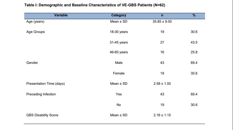
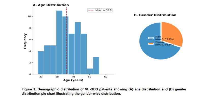
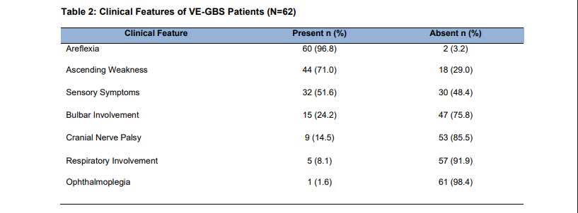
The male predominance (69.4%) with male:female ratio of 2.26:1. The mean presentation time was 2.58±1.00 days from symptom onset, with patients presenting on day 1 (n=9, 14.5%), day 2 (n=19, 30.6%), day 3 (n=21, 33.9%), and day 4 (n=13, 21.0%). History of preceding infection was present in 43 (69.4%) patients, with upper respiratory tract infection being more common (n=29, 67.4% of those with infection) than gastroenteritis (n=14, 32.6%). The mean GBS disability score at presentation was 2.16±1.15, indicating moderate disability. VE-GBS patients have clinical features, as summarized in Table II and depicted in Figure 2. The most common clinical observation was flexia, which was noted in 60 (96.8%) patients, which is in line with the diagnostic criteria of GBS. The traditional GBS presentation comprised of ascending pattern of weakness that occurred in 44 (71.0%) patients. Sensory symptoms such as paresthesias, numbness and tingling were reported in 32 (51.6%) patients. Bulbar involvement, which was experienced as dysphagia and dysarthria, was noted in 15 (24.2%) patients and cranial nerve palsy was noted in 9 (14.5%) patients. Close monitoring or intervention based on respiratory involvement was recorded in 5 (8.1%) patients. The least prevalent clinical finding was ophthalmoplegia that occurred in 1 (1.6%) patient.
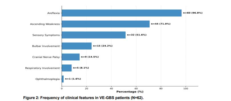
The horizontal bar chart demonstrates the frequency distribution of clinical manifestations, with areflexia (96.8%) being the most common presentation, followed by ascending weakness (71.0%) and sensory symptoms (51.6%).
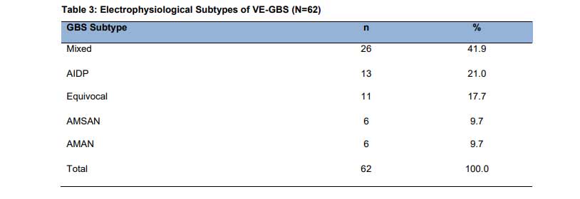
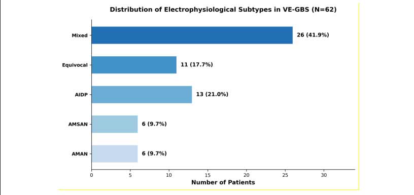
Table 3 shows the distribution of electrophysiological subtypes and Figure 3 illustrates it. The most common subtype was the mixed pattern, with demyelinating and axonal characteristics, and was seen in 26 (41.9%) patients. AIDP followed in 13 (21.0%) patients, equivocal pattern in 11 (17.7%) patients, AMSAN in 6 (9.7%) patients and AMAN in 6 (9.7%) patients. It is noteworthy that only 25 (40.3%) patients had classical GBS subtypes (AIDP, AMAN, AMSAN), and the rest 37 (59.7%) displayed mixed or equivocal cases that could not be classified.
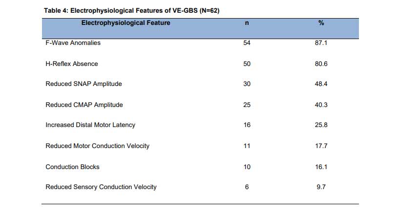
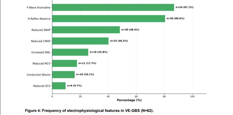
F-wave anomalies (87.1%) and H-reflex absence (80.6%) emerged as the most sensitive early diagnostic markers, detectable even when other nerve conduction parameters remained within normal limits.
Stratification analysis was performed to assess associations between GBS subtypes and demographic variables. Chi-square analysis revealed no statistically significant association between GBS subtypes and gender (χ²=5.683, df=4, p=0.224), age groups (χ²=6.368, df=8, p=0.606), presentation time (χ²=1.824, df=4, p=0.768), or history of preceding infection (χ²=2.515, df=4, p=0.642). These findings suggest that the distribution of electrophysiological patterns in VE-GBS is independent of demographic characteristics.
Discussion
This paper presents detailed information on clinical and electrophysiological features of Very Early Guillain-Barré Syndrome in a tertiary care Pakistani environment. Our results indicate that VE-GBS has characteristic patterns that are not similar to classical GBS presentations as assessed at advanced stages of disease, and have significant significance in early detection and decision-making in management.
Our demographic population revealed a mean age of 35.85 years with a strong male predominance (male to female ratio 2.26:1) which is similar to the international literature and other Asian-based epidemiological studies that reported a higher GBS incidence in males [3]. The male preponderance in our study is comparable to the 1.5-2:1 ratio in global epidemiological literature and other Asian studies [14]. The relatively young mean age of the population might be a result of the demographic distribution of our tertiary care center catchment area.
The clinically significant observation is that the prevalence of mixed (41.9) and equivocal (17.7) electrophysiological patterns is high, and only 40.3% of patients exhibit classical GBS subtypes. The result aligns with that of Berciano et al. who indicated that definitive classification into specific subtypes was only possible in 20-30% of VE-GBS [10]. The high percentage of mixed and equivocal patterns highlights the high diagnostic burden of very early presentations when electrophysiological abnormalities have not yet developed fully and have not been fully expressed.
Individual electrophysiological anomalies were found to be the most sensitive early signs of VE-GBS with F-wave (87.1) and H-reflex absence (80.6) being the most sensitive. This is in line with the results of the research conducted by Rasera et al., who also noted the absence of H-reflex in 73% and abnormal F-waves in 67% of early GBS cases [12]. The sensitivity of these parameters is high and may be attributed to the involvement of
the proximal nerve segments at early stages and this may be justified by the fact that the pathophysiology of GBS usually starts with inflammation at the level of nerve roots [15]. The implications of this finding are practical, because assessment of F-wave and H-reflex should be the first choice of clinicians in suspected VE-GBS. Areflexia (96.8%) and ascending weakness (71.0%) were found to be the cardinal features in our VE-GBS patients and are in line with the classical description of GBS [6].
Sensory symptoms were also found in 51.6 percent of the patients which is similar to the 60-65% reported in the international studies [16]. The respiratory involvement was reported in 8.1% of the patients, and it is necessary to carefully monitor respiratory conditions even in the early GBS manifestations [17]. No meaningful correlations between GBS subtypes and demographic factors indicate the possibility that the electrophysiological characteristics of VE-GBS can be closer to disease progression and development than to individual factors. This result has clinical implications, since it indicates that the initial electrophysiological classification would not be very reliable in determining the final disease phenotype [18]. Serial electrodiagnostic investigations can be required to identify materials classification of subtype.
There are a number of limitations of this study. The cross-sectional design did not allow determining the electrodiagnostic evolution with time and association with clinical outcomes. The single center design might not generalize to other settings and population. Moreover, we could not correlate electrophysiological subtypes and immunological profiles because of the lack of ganglioside antibody testing [19]. Further longitudinal research involving serial electrodiagnostic measures and antibody analysis would help in gaining a better understanding of how nerve conduction abnormalities change over time in VE-GBS and their prognosis [20].
Conclusion
VE-GBS among our population is mostly mixed or equivocal electrophysiologically and classical GBS subtypes can only be found in only 40.3% of cases in the first four days of symptoms. The most sensitive early electrophysiological parameters are F-wave abnormalities and H-reflex absence, both of which can be observed in more than 80% of patients in the presence of other normal parameters. The cardinal clinical characteristics are areflexia and ascending weakness. Identification of these patterns early together with clinical evaluation would help in the prompt diagnosis of the condition and ensure that the treatment commences in time which ultimately leads to better patient outcome with this potentially life threatening condition.
References
- Willison HJ, Jacobs BC, van Doorn PA. Guillain-Barré syndrome. Lancet. 2016;388(10045):717–727. https://doi.org/10.1016/S0140-6736(16)00339-1
- Sejvar JJ, Baughman AL, Wise M, Morgan OW. Population incidence of Guillain-Barré syndrome: a systematic review and meta-analysis. Neuroepidemiology. 2011;36(2):123–133. https://doi.org/10.1159/000324710
- McCombe PA, Hardy TA, Nona RJ, Greer JM. Sex differences in Guillain-Barré syndrome, chronic inflammatory demyelinating polyradiculoneuropathy and experimental autoimmune neuritis. Front Immunol. 2022;13:1038411. https://doi.org/10.3389/fimmu.2022.1038411
- Yuki N, Hartung HP. Guillain-Barré syndrome. N Engl J Med. 2012;366(24):2294–2304. https://doi.org/10.1056/NEJMra1114525
- Jacobs BC, Rothbarth PH, van der Meché FG, Herbrink P, Schmitz PI, de Klerk MA, et al. The spectrum of antecedent infections in Guillain-Barré syndrome. Neurology. 1998;51(4):1110–1115. https://doi.org/10.1212/WNL.51.4.1110
- Leonhard SE, Mandarakas MR, Gondim FAA, Batber K, Bber MR, Bber JK, et al. Diagnosis and management of Guillain-Barré syndrome in ten steps. Nat Rev Neurol. 2019;15(11):671–683. https://doi.org/10.1038/s41582-019-0250-9
- Hadden RD, Cornblath DR, Hughes RA, Zielasek J, Hartung HP, Toyka KV, et al. Electrophysiological classification of Guillain-Barré syndrome: clinical associations and outcome. Ann Neurol. 1998;44(5):780–788. https://doi.org/10.1002/ana.410440512
- Ho TW, Mishu B, Li CY, Gao CY, Cornblath DR, Griffin JW, et al. Guillain-Barré syndrome in northern China: relationship to Campylobacter jejuni infection and anti-glycolipid antibodies. Brain. 1995;118(Pt 3):597–605. https://doi.org/10.1093/brain/118.3.597
- Albertí MA, Alentorn A, Martínez-Yelamos S, Martínez-Matos JA, Povedano M, Saiz A, et al. Very early electrodiagnostic findings in Guillain-Barré syndrome. J Peripher Nerv Syst. 2011;16(2):136–142. https://doi.org/10.1111/j.1529-8027.2011.00344.x
- Berciano J, Orizaola P, Gallardo E, Pelayo-Negro AL, Sánchez-Juan P, Infante J, et al. Very early Guillain-Barré syndrome: a clinical-electrophysiological and ultrasonographic study. Clin Neurophysiol Pract. 2019;5:1–9. https://doi.org/10.1016/j.cnp.2019.10.002
- Uncini A, Kuwabara S. The electrodiagnosis of Guillain-Barré syndrome subtypes: where do we stand? Clin Neurophysiol. 2018;129(12):2586–2593. https://doi.org/10.1016/j.clinph.2018.09.025
- Rasera A, Romito S, Segatti A, Accorsi F, Squintani G, Tamburin S, et al. Very early and early neurophysiological abnormalities in Guillain-Barré syndrome: a 4-year retrospective study. Eur J Neurol. 2021;28(11):3768–3773. https://doi.org/10.1111/ene.14963
- Iqbal R, Asad MJ, Shah MB, Mahmood RT, Siddiqi S. Clinical and biochemical profile of Guillain-Barré syndrome in Pakistan. Neurosciences (Riyadh). 2021;26(3):242–247. https://doi.org/10.17712/nsj.2021.3.20200057
- Bhagat SK, Sidhant S, Bhatta M, Ghimire A, Shah B. Clinical profile, functional outcome, and mortality of Guillain-Barré syndrome: a five-year tertiary care experience from Nepal. Neurol Res Int. 2019;2019:3867946. https://doi.org/10.1155/2019/3867946
- Gupta D, Nair M, Baheti NN, Sarma PS, Kuruvilla A. Electrodiagnostic and clinical aspects of Guillain-Barré syndrome: an analysis of 142 cases. J Clin Neuromuscul Dis. 2008;10(2):42–51. https://doi.org/10.1097/CND.0b013e31815c0b6a
- Akbayram S, Doğan M, Akgün C, Peker E, Sayin R, Aktar F, et al. Clinical features and prognosis with Guillain-Barré syndrome. Ann Indian Acad Neurol. 2011;14(2):98–102. https://doi.org/10.4103/0972-2327.82794
- Orlikowski D, Sharshar T, Porcher R, Annane D, Raphael JC, Clair B. Prognosis and risk factors of early onset pneumonia in ventilated patients with Guillain-Barré syndrome. Intensive Care Med. 2006;32(12):1962–1969. https://doi.org/10.1007/s00134-006-0426-7
- Fokke C, van den Berg B, Drenthen J, Walgaard C, van Doorn PA, Jacobs BC. Diagnosis of Guillain-Barré syndrome and validation of Brighton criteria. Brain. 2014;137(Pt 1):33–43. https://doi.org/10.1093/brain/awt285
- Kuwabara S, Yuki N. Axonal Guillain-Barré syndrome: concepts and controversies. Lancet Neurol. 2013;12(12):1180–1188. https://doi.org/10.1016/S1474-4422(13)70215-1
- Doets AY, Verboon C, van den Berg B, Harbo T, Cornblath DR, Willison HJ, et al. Regional variation of Guillain-Barré syndrome. Brain. 2018;141(10):2866–2877. https://doi.org/10.1093/brain/awy232