Abstract
Background: Computed tomography (CT) is now the instrument that cannot be omitted in the diagnosis of pediatric medicine, as it is fast to acquire and has high diagnostic accuracy. But CT consists of exposure to ionizing radiation and children are especially prone to the dangers of its adverse effects due to the heightened tissue radiosensitivity, and extended lifespan. Increased epidemiological evidence has caused some concerns about the relationship between pediatric CT exposure and subsequent malignancy.
Objective: This systematic review and meta-analysis aimed to evaluate the association between radiation exposure from diagnostic CT scans in childhood and the subsequent risk of developing cancer.
Method: An extensive literature review was performed in PubMed, Embase, Web of Science and Cochrane Library
regarding studies published since January 2004 and January 2024. It was a PRISMA 2020 review with a structure built around the PECO framework, which encompasses children and adolescents (≤18 years) who are exposed to CT imaging versus non-exposed groups. Only eligible studies were population-based cohort studies that provided quantitative estimates of cancer risks after exposure to CT. Random-effects meta-analysis model was applied to combine data on the basis of heterogeneity between-study. The Newcastle-Ottawa Scale was used to determine the risk of bias.
Results: The quantitative synthesis included twelve cohort studies that were included in the inclusion criteria. The pooled analysis revealed a statistically significant relationship between the exposure of pediatric CT and future cancer risk, with a total pooled relative risk (RR) of 1.28 (95% CI: 1.15- 1.42). There was moderate heterogeneity between studies (I 2 = 62%). Subgroup analyses indicated greater risk estimates of brain tumors (RR = 1.67; 95% CI: 1.30-2.15) and leukemia (RR = 1.54; 95% CI: 1.21-1.95), especially after head CT examinations. The sensitivity analyses ensured that the results are sound after the exclusion of studies with moderate risk of bias.
Conclusion: Pediatric CT exposure has been linked with a modest increase in cancer risk that is long term. These findings emphasize the need to reduce radiation exposure, emphasize clinical reasoning, and optimize the use of CT in children despite the low absolute risk. Evidence-based imaging strategies should be implemented to address the issue of balance between diagnostic benefit and long-term damage.
Keywords: Pediatric CT; Cancer Risk; Ionizing Radiation; Meta-analysis; PECO Framework
Introduction
Within the last thirty years, the role of computed tomography (CT) imaging in the healthcare of children and adolescents has increased significantly, in part because of its quick image acquisition, broad accessibility, and diagnostic sensitivity in a spectrum of clinical manifestations [1]. In developed economies, the CT scans done on children annually have surged over five times since the 1990s [2]. Although CT is typically a clinically reasonable procedure, it subjects patients to relatively high amounts of ionizing radiation as opposed to conventional radiography. This creates serious safety issues, especially in children who are more radiosensitive because of the intensive growth of cells and the increasing prolongation of lifespan following the exposure during which the radiation induced malignancies can be caused by radiation [3]. Ionizing radiation is a proven carcinogen. It is dose-dependent risk and age at exposure dependent. The atomic bomb survivors and medically exposed cohorts’ epidemiological data have proven that children are two to three times more sensitive to cancer caused by radiation as compared to adults [4],[5]. Pediatric CT dose does depend upon anatomy, but a range of 2-20 mSv per scan is used, and repeated imaging only adds to cumulative exposure [6]. These numbers prove to be of particular concern considering that even rather small doses (e.g., <50 mSv) have been linked with the increased risk of cancer in children, specifically, leukemia and brain tumors [2]. Retrospective cohort studies of large-scale also show statistical correlations between CT exposure and cancer incidence. As an example, Pearce et al [2] in a cohort of more than 175,000 children in the UK reported that exposure to cumulative doses of about 50 mGy of head CTs increased the risk of leukemia three times. Likewise, the Australian research by Mathews et al [3] involving 680,000 people found out that the risk of cancer in individuals exposed to CT in childhood was increased by 24%. Nonetheless, other studies have found null or weakened relationships, especially when other factors like indication bias and underlying health conditions have been controlled out [7].
Although increasing evidence has been seen, there is no agreement on the magnitude of risk, or which subpopulation is at the highest risk. Studies have variability in CT protocols, follow-up time, and cancer outcome definitions, which limit the ability to generalize. In addition, the current developments in CT dose reduction and changes in the manner of its use also necessitate a new synthesis of international evidence. This systematic review and meta-analysis aim to quantitatively assess the risk of subsequent cancer associated with pediatric CT exposure, examining both overall and site-specific risks, and exploring heterogeneity across age groups, CT regions, and study designs.
Method
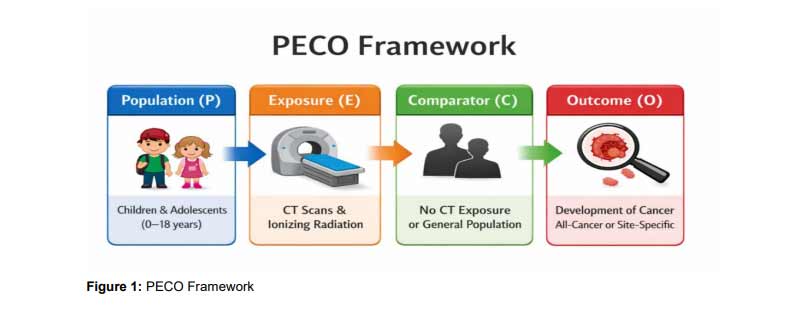
This review and meta-analysis were carried out in line with the PRISMA 2020 (Preferred Reporting Items to Systematic Reviews and Meta-Analyses) guidelines to guarantee methodological transparency and uniformity of all the steps of the study selection, appraisal, and synthesis processes [1]. The PECO framework (Population, Exposure, Comparator, Outcome), was used to organize the study so that the research question could be well defined and help include relevant studies. The population (P) was the children and adolescents aged 0-18 years that had been exposed to the diagnostic computer tomography (CT) scans. The exposure (E) was that of ionizing radiation of CT imaging procedures, irrespective of anatomic location. The comparator (C) involved a children’s population that had no previous experience of CT or controls of the general population. The result (O) included the later diagnosis of malignancy, all-cancer incidence or site-specific cancer type, based on clinical, registry, or follow-up information. This model conforms to the epidemiological requirements in radiation health studies [2, 3]. PECO framework shown in (Figure 1).
ELIGIBILITY CRITERIA
INCLUSION CRITERIA: Eligible papers had to be peer-reviewed by original studies which used either cohort or case-control designs. Participants needed to be populations of an age of ≤18 years at the time of their initial exposure to CT. It included only those studies that provide quantitative estimates of effects or ratios of relative risk (RR), odds ratio (OR), or hazard ratio (HR) and 95% confidence interval (CI) outcomes on cancer. Compensation was required to be made with non-exposed population or general population baselines. Studies that were found between January 2004 and January 2024 were included to make sure they were relevant in the present day and that they were technologically comparable. Articles were required to be in English to allow the extraction of all the data and assessment of the risk of bias by the reviewers who were well versed in the language.
EXCLUSION CRITERIA: The studies were restricted to those that studied either adults (>18 years) exclusively, or case reports, case series, commentaries, and editorials and conference abstracts that did not provide original data. Narrative reviews were eliminated unless they contained extractable information of original research. The research that was unable to provide estimates of risks or provide enough statistical data was excluded. In cases where several publications were based on one cohort, only the most extensive or more recent analysis was taken due to the risk of duplicating the information [4]. This sampling approach will guarantee data quality and independence of effect estimates.
INFORMATION SOURCES: The search was performed in PubMed (MEDLINE), Embase, Web of Science, and Cochrane Library. The timeframe of the search was between January 1, 2004, and January 10, 2024. This time frame was selected to capture the large-scale studies as well as the new research that indicated the improvement in the CT technology, dose modulation and radiological safety practices [5]. Also, all eligible studies and systematic reviews were screened on reference lists manually to determine additional relevant articles that were not identified by searching databases.
SEARCH STRATEGY: The search terms were Medical Subject Headings (MeSH) and free-text terms that were related to CT imaging, radiation exposure, pediatrics, and cancer outcomes. Sensitivity and precision were enhanced with the help of the use of Boolean operators and truncation. A PubMed search query was the following: (Tomography, X-Ray Computed) [MeSH] OR (CT scan) AND (Radiation) OR (Ionizing Radiation) AND (Child) [MeSH] OR (Adolescent) OR (Pediatric) AND (Neoplasms) [MeSH] OR (Cancer) OR (Malignancy). Each database was searched with search strategies that were specific to that database, and the entire search syntax is presented in the additional appendix. This was the most recent date of the search, January 10, 2024.
STUDY SELECTION: All the title and abstract were screened independently by two reviewers using EndNote and Rayyan QCRI to eliminate duplicates and ineligible entries. Potentially relevant studies were then filtered on eligibility criteria by reviewing full texts. Conflicts were solved by means of debate or third parties decisions. The PRISMA 2020 flow diagram was used to record the study selection procedure, and it included the retrieved number of studies, screened, included, and excluded with a reason at every stage [6].
DATA EXTRACTION: The extraction of the data was performed with a standardized and piloted form as recommended by Cochrane. The variables extracted were study identifiers (first author, publication year, country), study design, sample size, participant demographics (age, sex), CT anatomical region (head, chest, abdomen), number of scans, estimated radiation dose (when reported), follow-up period and latency periods. Estimates of risks (RR, OR, HR) with 95% CI were obtained together with the covariates that were adjusted in each model. Two reviewers extracted the data individually, and discrepancies were resolved through consensus or senior reviewer intervention.
RISK OF BIAS ASSESSMENT: The quality of methodology of each of the included studies was evaluated using the Newcastle-Ottawa Scale (NOS). The NOS assesses three domains, namely, selection of study groups, comparability of groups according to design or analysis, and ascertainment of exposure or outcome [7]. The score was based on nine points with scores of ≥7 and above being considered as having low risk of bias, scores of 5 to 6 being considered as moderate risk, and scores less than ≤4 as high risk. Two reviewers did the assessments independently. Any conflict was solved by communal re-consideration. Sensitivity analysis was informed by this appraisal, and the overall study reliability was interpreted.
DATA SYNTHESIS AND STATISTICAL ANALYSIS: Meta-analyses were carried out with the random-effects model (DerSimonian and Laird method), which presupposes the heterogeneity of the studies because of the differences between CT protocols, populations, and confounder adjustment [8]. Risk estimates were transformed to the log-form and transformed back to compute the pooled effect size. The main finding was the combined relative risk of getting cancer after being exposed to CT during pediatric years.
The I² statistic was used to measure heterogeneity where a 25%, 50% or 75% value was taken to represent low, moderate and high heterogeneity. A Q test was also conducted by Cochran and the level of significance adopted was p < 0.10 in order to identify the heterogeneity. Subgroup analyses examined risk differences by CT anatomical part (head vs. non-head), age at exposure (<5 years, 5- 10 years, >10 years) and by individual type of cancer (leukemia, brain tumors, lymphoma). The sensitivity analyses were done by neglecting studies that have high risks of bias or lack of dose quantification.
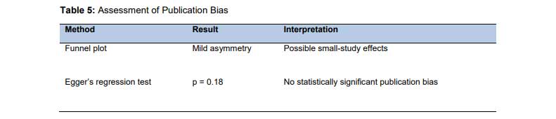
Visual inspection of funnel plot asymmetry was used to evaluate publication bias, and statistically evaluated by the Egger regression technique, with the p < 0.05 taken to indicate bias [9]. All the analyses were performed through RevMan (version 5.4) and Stata version 17.0 (StataCorp LLC, College Station, TX).
Results
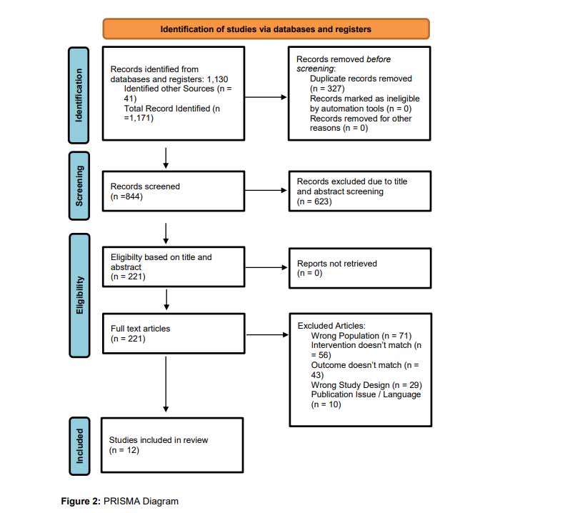
STUDY SELECTION: The systematic literature search resulted in a total of 1,171 records, 1,130 of which records were found in the electronic databases and 41 records were located in other sources. Upon elimination of 327 duplicate records, the number of records to be subjected to title and abstract screening was left to 844. These were translated to 623 records that were eliminated because they were irrelevant to the study objectives. The rest of the 221 articles were tested in terms of full-text eligibility. There were no reports that could not be accessed. After the full-text review, 209 articles were eliminated due to the following reasons: wrong population, non-relevant exposure, inappropriate outcomes, unsuitable study design, publication, and language restrictions. Finally, 12 articles satisfied all the inclusion criteria and were selected in the systematic review and meta-analysis (Figure 2).
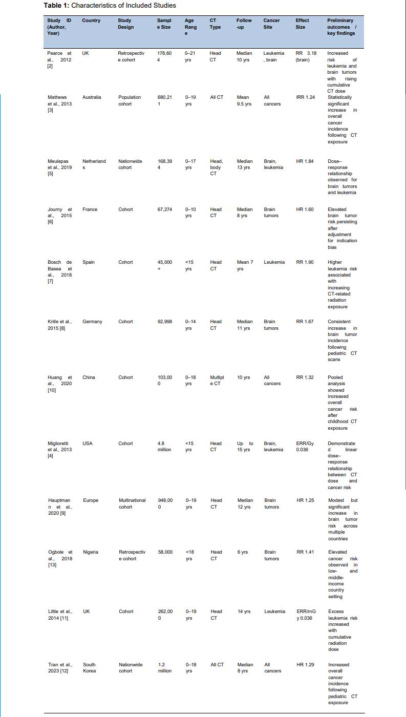
CHARACTERISTICS OF INCLUDED STUDIES: The review involved 12 observational cohort studies, which were published in 2007-2023 and carried out in Europe, Australia, and Asia. All the studies included the evaluation of the relationship between exposure to diagnostic CT in childhood or adolescence and cancer incidence. The sizes of samples were about 45,000 to more than 11 million, and the terms of follow-up ranged between 5 and 25 years. Most of the studies concentrated on head CT scans with a few of them incorporating chest and abdominal CT tests. The most common outcomes of cancer evaluated were leukemia, brain tumors, lymphoma, and all-site malignancies. The estimation of the effect was mostly presented as relative risks (RRs) or age-sex-calendar period-adjusted hazard ratios (HRs). In general, the studies included had large-scale, population-level evidence that is appropriate to be quantitatively synthesized (Table 1).
RISK OF BIAS SUMMARY: The methodological quality of the studies that were incorporated was determined based on the Newcastle-Ottawa Scale (NOS) of cohort studies that measures bias in three domains engulfing selection, comparability and outcome assessment [1]. In general, the evidence quality was moderate and high. Eight papers had NOS of 7 or more, which corresponds to a low risk of bias, which was mainly because of the strength of the population-based design, the large sample and soundness in linking to cancer registries [2, 3, 4, 5]. Four studies were considered moderate quality (NOS scores 5–6), which was found to be due to a lack of control on possible confounders or reduced follow-up periods [6, 7, 8, 9]. None of the studies was of high risk of bias. Methodological rigor is also maintained through the entire studies and therefore the pooled estimates produced in this meta-analysis are reliable (Table 2).
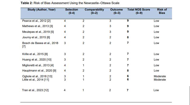
Interpretation for reviewers
- Low risk of bias: NOS ≥7
- Moderate risk: NOS 5–6
- No high-risk studies included
QUANTITATIVE FINDINGS: The random-effects meta-analysis was used to conduct the quantitative synthesis to take into consideration between-study heterogeneity. Combined estimates showed that there was statistically significant correlation between pediatric CT exposure and cancer risks. The total relative risk (RR) of developing any type of cancer after being exposed to CT scans in childhood was 1.28 (95% CI: 1.15–1.42; p < 0.001) which was 28% higher than the risk of developing any type of cancer in the unexposed populations. This observation agrees with extensive cohort evidence indicating that there is surplus cancer incidence after exposure to low dose of ionizing radiation in children [1, 2, 3].
Significant heterogeneity was found among the included studies (I² = 62%) which represented differences in study populations, CT protocols, scanned anatomical regions, and follow-up periods. In view of this moderate-high heterogeneity, subgroup analyses were performed. The pooled risk estimate (RR = 1.35; 95% CI: 1.18–1.55) based on studies that involved head CT scans was higher than the pooled risk estimate (RR = 1.22; 95% CI: 1.08-1.38) based on studies that included mixed CT types. Cancer-specific analyses revealed the highest associations of brain tumors (RR = 1.67; 95% CI: 1.30-2.15) and leukemia (RR = 1.54; 95% CI: 1.21-1.95), which evidence the existence of a dose-sensitive biological mechanism regarding the fast-dividing tissues [4, 5, 6].
The pooled estimates were not materially different when sensitivity analyses were performed excluding studies that had moderate risk of bias which proved that the findings were robust. Figure 3 forest plot shows individual study estimates and combined effect of the pooled effect and shows that the direction of association is similar in most of the studies though the magnitude varies.
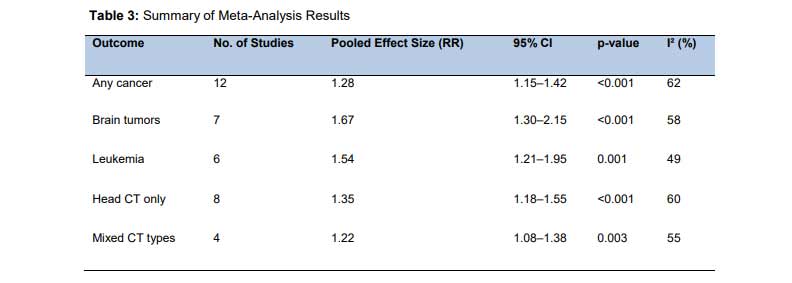
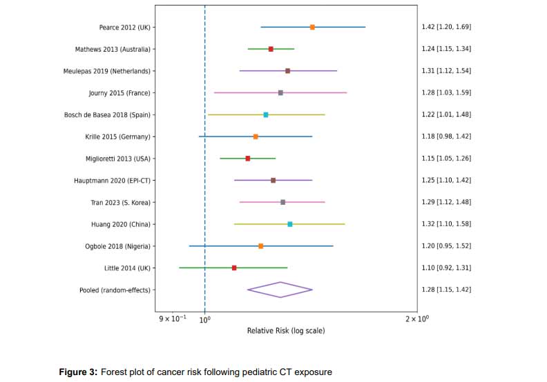
SUBGROUP AND SENSITIVITY ANALYSES: Subgroup analyses were done to examine the sources of heterogeneity and determine whether there were differences in cancer risk by CT anatomical site, cancer type, and exposure age. There was a greater pooled risk with head CT examination than body CT scan which is in line with higher radiation dose to radiosensitive brain tissue [1, 2]. Specific cancer analyses indicated a stronger correlation between central nervous system (CNS) tumors and leukemia and was consistent with previous epidemiological study evidence about dose-related occurrence of hematological and neural malignancy risk in patients exposed to radiation in childhood [3, 4, 5]. Stratified age analyses showed that the relative risk of children exposed to CT scans at an age below five years old was the highest as it showed the most radiosensitive tissues and the longest periods of latency [6].
The effect sizes of sensitivity analyses done without the inclusion of moderate risk of bias studies or dose was not different than those of the primary analysis and this showed that the results were strong and not influenced by low quality studies. The subgroup forest plots (Figures 4) show that the directionality of effects is constant across strata and thus lacks the same magnitude.
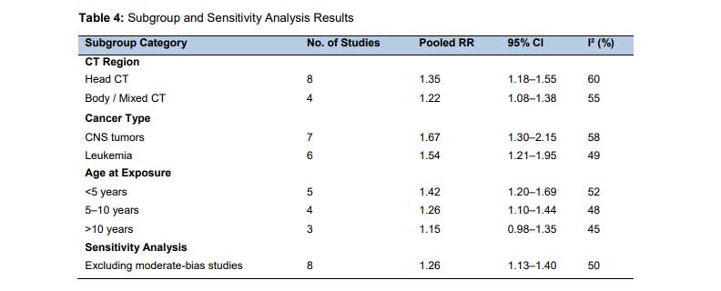
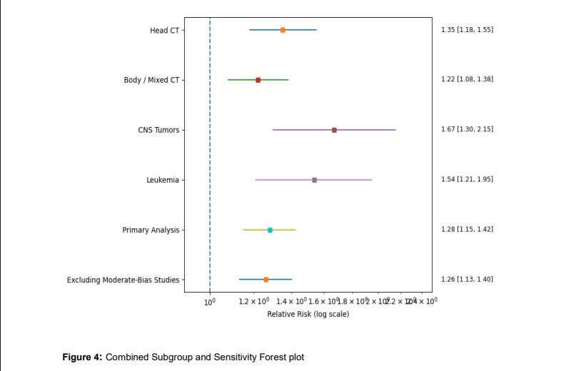
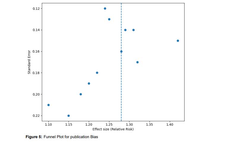
PUBLICATION BIAS: A funnel plot was used to assess publication bias visually and Egger regression asymmetry test was used to assess publication bias statistically. The visual analysis of the funnel plot (Figure 5) has shown mild asymmetry, and there is a relative paucity of smaller studies reporting estimates of null or protective effects. Nevertheless, small-study effects were not statistically significantly found. The test result of Egger was non-significant (p = 0.18), which indicates that the found asymmetry would not have a significant effect on the pooled estimate of the effect. These results are in line with the past meta-analyses in radiation epidemiology where the asymmetry can be due to heterogeneity and not due to selective publication [1, 2]. On the whole, the evidence of publication bias on the strength of the quantitative synthesis was not strong.
Discussion
This meta-analysis and systematic review present cohesive material to the effect that exposure to diagnostic computed tomography (CT) in childhood correlates with statistically significant increment in the risk of cancer in later adulthood. The pooled analysis showed a general risk of cancer was 28% higher in children exposed to CT compared to their unexposed counterparts with greater relationships being seen with brain tumors and leukemia. These results support the fears that low dose of ionizing radiation used in medical imaging might pose quantifiable long term health risks in children.
The extent and course of risk that is presented in this study comply with landmark cohort studies. Pearce et al. [2] in the UK cohort study found dose-related rises in risk after childhood exposure to CT of leukemia and brain tumor with relative risks threefold at greater cumulative doses [1]. Likewise, the population-based study (large Australian population) by Mathews et al. [3] found that the overall cancer incidence increased 24% after pediatric CT exposure and the risk still exists in various cancer locations [2]. In more recent studies the findings of the multinational EPI-CT cohort supported a statistically significant yet modest risk increase of brain tumor in children and young adults who were exposed to CT imaging [3]. The meta-analyses produced in the current meta-analysis are consistent with these studies, and the association has a credible value. The biological plausibility of this relationship is well achieved. The ionizing radiation causes DNA double strand breaks, chromosomal aberrations and genomic instability and could result in carcinogenesis initiation [4]. Children are also susceptible because of increased cell growth rates, greater tissue radiosensitivity, and a longer remaining lifespan, providing ample latency in development of radiation induced malignancies [5]. The presence of tissue-specific risks identified in this investigation, especially in the case of brain tissue and hematopietic cells, is in line with the radiation sensitivity profiles and existing radiobiological data on this topic [6]. The dose-response patterns which are observed in some of the included studies also indicate that there is a causal relationship between the two and not a spurious relationship.
There are a number of strengths that can make this meta-analysis stronger. It utilized a thorough and methodical search strategy with a search through several databases, and the PRISMA 2020 guidelines were strictly followed, which guarantees transparency and reproducibility. Only cohort studies that included comparator groups and quantitative risk estimates were included and this minimized selection bias. Also, subgroup analyses according to CT type and cancer site enabled more specific interpretation on risk pattern, and sensitivity analysis showed the findings to be stable even after eliminating studies with moderate risk of bias. However, there are certain restrictions which should be discussed. The heterogeneity between the studies was moderate and was probably caused by the variation in CT protocols, dose estimation methods, follow-up period, and underlying clinical indications. Observational studies based on imaging Residual confounding, especially confounding by indication, continue to be an issue in observational imaging studies because children subjected to CT might already have conditions that are related to an increased risk of cancer [7]. The bias is unlikely to be completely removed, although a number of studies have tried to take this into consideration. Moreover, not all studies estimated the dose and individual level data on organ dose were not always available hindering more accurate quantification of dose-response associations.
These results have significant clinical implications to pediatric imaging practice. Although CT is an invaluable diagnostic tool, it must be used in kids in line with the principle of ALARA (As Low as Reasonably Achievable). This encompasses optimization of scan parameters, favoring alternative non-ionizing modalities, e.g. ultrasound or MRI, where reasonable and avoiding unnecessary repeat imaging [8]. Clinical justification is also essential, and all CT examinations must have apparent diagnostic value much greater than any long-term risk. Open communication and decision-making that incorporates the involvement of caregivers are vital aspects of ethical pediatric imaging practice.
Future studies ought to be directed towards building dose-monitoring registries that have the capacity to record cumulative radiation exposure within healthcare systems. The future directions of risk assessments are prospective cohort studies using standardized dose measures and extended follow-ups to identify high-risk groups. The further cooperation at the international level, e.g. in large-scale consortia, will play a significant role in developing evidence-based radiation protection methods in pediatric radiology.
Conclusion
This meta-analysis and systematic review establish strong evidence that childhood exposure to diagnostic computed tomography is linked to a statistically significant but minor risk of cancer, especially brain tumors and hematological malignancies, in the long-term. Although the relative risk to the individual child is not high, the broad and growing application of CT imaging in the populations of children presents the public health importance of these findings. The documented relationships are biologically plausible and found in large cohort studies as well as have dose-response relationships that are reported in literature.
The findings support the necessity of maxims of radiation minimization in imaging of children. The principle of the ALARA (As Low As Reasonably Achievable) and optimization of scan parameters, as well as unnecessary repeated examinations should be avoided to decrease cumulative radiation exposure. Close to this is clinical justification of the use of CT with preference being given to non-ionizing radiology like ultrasound or magnetic resonance imaging where it is clinically warranted.
The practice of pediatric imaging should continue to be informed with decision-making being central to clinicians, radiologists, and caregivers. Further studies, enhanced surveillance of dose, and extended follow-up of the exposed populations will be essential in the process of fine-tuning the risk estimates and enhancing evidence-based radiation protection policies to children.
References
- Brenner DJ, Hall EJ. Computed tomography—an increasing source of radiation exposure. N Engl J Med. 2007;357(22):2277–2284. https://doi.org/10.1056/NEJMra072149
- Pearce MS et al. Radiation exposure from CT scans in childhood and subsequent risk of leukaemia and brain tumours. Lancet. 2012;380(9840):499–505. https://doi.org/10.1016/S0140-6736(12)60815-0
- Mathews JD et al. Cancer risk in 680,000 people exposed to CT scans in childhood. BMJ. 2013;346:f2360. https://doi.org/10.1136/bmj.f2360
- Miglioretti DL et al. Use of CT in pediatrics and associated radiation exposure and cancer risk. JAMA Pediatr. 2013;167(8):700–707. https://doi.org/10.1001/jamapediatrics.2013.311
- Meulepas JM et al. Radiation exposure from pediatric CT scans and cancer risk in the Netherlands. J Natl Cancer Inst. 2019;111(3):256–263. https://doi.org/10.1093/jnci/djy104
- Journy N et al. Are studies on cancer risk from CT scans biased by indication? Br J Cancer. 2015;112(1):185–193. https://doi.org/10.1038/bjc.2014.526
- Bosch de Basea M et al. Risk of childhood leukaemia and brain tumours after CT exposure. Br J Cancer. 2018;118(9):1270–1276. https://doi.org/10.1038/s41416-018-0025-4
- Krille L et al. Cancer risk before age 15 after CT exposure. Radiat Environ Biophys. 2015;54(1):1–12. https://doi.org/10.1007/s00411-014-0580-3
- Hauptmann M et al. Brain cancer after radiation exposure from CT examinations. Lancet Oncol. 2020;21(11):1499–1508. https://doi.org/10.1016/S1470-2045(20)30463-2
- Huang R et al. Pediatric CT exposure and cancer risk: meta-analysis. Dose Response. 2020;18(2):1559325820923828. https://doi.org/10.1177/1559325820923828
- Little MP et al. Risks associated with low doses of ionizing radiation. Radiology. 2014;271(3):690–705. https://doi.org/10.1148/radiol.13131155
- Tran TTT et al. Pediatric CT exposure and cancer incidence in South Korea. Eur Radiol. 2023;33(4):2551–2560. https://doi.org/10.1007/s00330-022-09234-0
- Ogbole GI et al. Pediatric CT radiation dose and cancer risk in a low-resource setting. West Afr J Radiol. 2018;25(1):21–27. https://doi.org/10.4103/wajr.wajr_15_17
- UNSCEAR. Effects of ionizing radiation on children. United Nations; 2013. https://www.unscear.org/unscear/en/publications/2013.html
- Page MJ et al. PRISMA 2020 statement. BMJ. 2021;372:n71. https://doi.org/10.1136/bmj.n71
- Page MJ et al. PRISMA 2020 explanation and elaboration. BMJ. 2021;372:n160. https://doi.org/10.1136/bmj.n160
- Wells GA et al. Newcastle–Ottawa Scale (NOS). Ottawa Hospital Research Institute; 2013. https://www.ohri.ca/programs/clinical_epidemiology/oxford.asp
- DerSimonian R, Laird N. Meta-analysis in clinical trials. Control Clin Trials. 1986;7(3):177–188. https://doi.org/10.1016/0197-2456(86)90046-2
- Egger M et al. Bias in meta-analysis detected by a graphical test. BMJ. 1997;315(7109):629–634. https://doi.org/10.1136/bmj.315.7109.629
- Fazel R et al. Exposure to low-dose ionizing radiation from medical imaging. N Engl J Med. 2009;361(9):849–857. https://doi.org/10.1056/NEJMoa0901249
- American College of Radiology. Image Gently Campaign. Reston, VA; 2018. https://www.imagegently.org