
Clinical Profile and Predictors of Hepatic Encephalopathy in Liver Cirrhosis: A Cross-Sectional Study at Mardan Medical Complex, Mardan
Correspondence and article notes 8
ABSTRACT
Background: A common and dangerous complication of liver cirrhosis, hepatic encephalopathy (HE) dramatically raises hospitalization, morbidity, and mortality. There is limited local data exist from LMICs. Among hospitalized patients with cirrhosis at Mardan Medical Complex, Mardan.
Objective: The goal is to ascertain the prevalence, grade distribution, precipitating events, and independent predictors of early HE (within 48 hours).
Methods: Between January and August of 2025, a prospective cross-sectional study was carried out. Adults who had cirrhosis and were consecutive were enrolled. At admission and 48 hours later, HE was evaluated using the West Haven criteria. Clinical and analytical parameters were collected, and precipitating factors were systematically screened. Using multivariable logistic regression with bootstrap validation, independent predictors were found. AUC and calibration measures were used to assess the model's performance, and a bedside nomogram was produced.
Results: 256 patients (mean age 54.3 ± 11.8 years; 59.8% male) were included out of 281 screened. 39.5% (95%CI 33.7–45.6) had HE, and 81.2% had Grade I–II. The most common precipitants were infection (36.6%) and hyponatremia (29.7%). Independent predictors included prior HE (AOR 2.41, 95% CI 1.34–4.33), infection (AOR 2.09, 1.28–3.43), lower sodium (per 3 mmol/L decrease: AOR 1.35, 1.16–1.57), higher creatinine (per 0.5 mg/dL increase: AOR 1.28, 1.04–1.58), higher bilirubin (per 1 mg/dL increase: AOR 1.06, 1.02–1.10), higher INR (per 0.5 increase: AOR 1.31, 1.06–1.62), constipation (AOR 1.68, 1.02–2.77), and exposure to psychoactive drugs (AOR 1.95, 1.06–3.57). Excessive diuretics showed borderline significance (AOR 1.72, 0.99–2.98; p = 0.054). The model demonstrated high calibration and strong discrimination (AUC 0.83).
Conclusion: Modifiable factors play a major role in the prevalence of early HE in cirrhosis. In environments with limited resources, the validated bedside nomogram may facilitate early triage and focused interventions.
Keywords: Hepatic Encephalopathy; Liver Cirrhosis; Risk Factors; Nomograms; Hyponatremia; Pakistan
INTRODUCTION
One of the most common and serious complications of liver cirrhosis is hepatic encephalopathy (HE), which significantly increases morbidity, mortality, and healthcare use globally [1,2]. About 30 to 45 percent of cirrhosis patients have it at some point throughout their illness, and it is the primary cause of hospitalizations for liver-related conditions [3, 4]. Reversible causes include illness, electrolyte imbalances, gastrointestinal hemorrhage, constipation, dehydration, or sedative exposure are frequently the cause of HE, which can range clinically from mild cognitive impairment to severe coma [2,5].
Cirrhosis is one of the leading causes of years of life lost globally, and the burden of liver disease is still increasing [6]. While viral hepatitis is still quite common in low- and middle-income nations, epidemiologic shifts are moving the disease etiology away from viral hepatitis and toward metabolic dysfunction–associated steatotic liver disease (MASLD) [6,7]. The risk of HE as a decompensating event is increased by this dual burden. Persistent hepatitis C virus (HCV) infection and rising MASLD incidence are the main causes of Pakistan being amongst the highest rates of chronic liver disease in the area [8–10]. About 6% of the general population is thought to have HCV seroprevalence [8], whereas about one-third of adults have MASLD [9, 10]. This puts a lot of people at risk for HE and decompensated cirrhosis, especially in provinces with minimal resources like Khyber Pakhtunkhwa.
According to international standards, HE-related morbidity and hospital resource utilization can be considerably decreased by early detection of precipitating variables and systematic care [2,5,11]. Early risk stratification can be facilitated by clinical prediction models and organized bedside methods, but there is still a dearth of data in South Asia on this topic, particularly with regard to early inpatient HE (on admission and within the first 48 hours) [12].
Although Pakistan has a high rate of cirrhosis admissions, there is a dearth of prospective data on the incidence of HE, its contributing causes, and its independent predictors at presentation. In environments with limited resources, identifying locally relevant predictors can assist in customizing triage, monitoring, and bundled care.
The aim of this study was to prospectively assess adults with cirrhosis who were admitted to a tertiary hospital in Mardan, Pakistan, in order to determine the grade distribution and prevalence of hepatic encephalopathy at presentation or within the first 48 hours of hospitalization, describe common precipitating factors, find independent predictors of HE using multivariable logistic regression with internal validation, and convert the significant predictors into a nomogram that can be used in clinical settings for early risk assessment.
Methods
This prospective cross-sectional study was conducted in Department of Gastroenterology at Mardan Medical Complex, Mardan, a tertiary-care teaching hospital with 24-hour emergency, laboratory, endoscopic, and radiology facilities. January 2025 to August 2025 was the study's time frame; setup took one month, enrollment took six months, and data cleaning and analysis took two months. During this time, consecutive eligible patients from the inpatient and medical emergency wards were included. Adult patients having a confirmed diagnosis of liver cirrhosis were included in the target population. A hepatologist's prior documentation, endoscopic or radiologic evidence of portal hypertension, such as varices or portosystemic collaterals, transient elastography ≥14 kPa when available, or characteristic imaging findings of cirrhosis or portal hypertension (nodular liver, splenomegaly, collaterals) were all considered criteria for defining cirrhosis. Histology wasaccepted provided it was included in the medical file.
Clinically, hepatic encephalopathy was classified using the West Haven criteria, which ranged from 0 to IV. [2, 13] To determine the presence and severity of HE, patients were evaluated within 24 hours of presentation and again 48 hours later. HE was defined as Grade ≥1 at presentation or within 48 hours for the purposes of the primary analysis. Sleep-wake disruptions, psychomotor abnormalities, asterixis, and consciousness level were recorded. When appropriate, a quick Number Connection Test A was employed to aid in early grade classification. [14] A standardized checklist was used to systematically screen for common precipitating factors, including infection (including spontaneous bacterial peritonitis, urinary tract infection, or pneumonia), electrolyte disorders (hyponatremia or hypokalemia), constipation, dehydration or excessive use of diuretics, use of sedatives or psychoactive drugs, recent protein load, acute blood loss, acute kidney injury, and nonadherence to lactulose. [2]
All patients with proven cirrhosis who presented throughout the recruitment period and were at least 18 years old were eligible. Exclusion criteria included severe hypoglycemia unrelated to cirrhosis, postictal states, alcohol intoxication or withdrawal mimicking HE, severe psychiatric illness interfering with assessment, primary neurologic disorders explaining altered mentation, and recent procedural anesthesia confounding mental status. To reduce selection bias, consecutive sampling was employed.
The single proportion formula was used to estimate the sample size. The necessary minimum sample size was roughly 196 patients for a 95% confidence level (Z = 1.96), a conservative predicted prevalence (p = 0.50) of hepatic encephalopathy among hospitalized cirrhotic patients based on previous literature, and a 7% absolute precision (d = 0.07). Assuming a HE prevalence of roughly 35%, the necessary total grew to 229 in order to permit multivariable logistic regression with up to eight predictors and guarantee ≥10 events per variable. The ultimate target sample size was set at 260 participants after taking into consideration about 10% of the data was incomplete [3]
Data on demographics, the etiology of cirrhosis (hepatitis B, hepatitis C, MASLD/NAFLD, alcohol, autoimmune, cholestatic, and cryptogenic), the length of the illness, previous decompensations, such as ascites or HE, medication history, physical examination, including West Haven grading and ascites assessment, and laboratory tests conducted within 24 hours of admission (CBC, PT/INR, bilirubin, transaminases, albumin, creatinine, sodium, potassium, glucose, CRP, and ascitic fluid analysis when necessary) were all gathered using a structured proforma. MELD-Na and Child-Pugh scores were computed. The presence or absence of precipitating variables was noted. During a pilot phase, all evaluations were carried out by qualified medical officers who had inter-rater calibration. Standardized criteria, HE grading training, duplicate data entry, and predetermined analytical techniques all helped to reduce bias. Selection bias was lessened by consecutive enrollment. Information bias was reduced by using initial labs and standardizing proforma-based ascertainment. Multivariable modeling helped to mitigate confounding. Complete-case analysis was utilized for missing data with a percentage of less than 5%; multiple imputation was employed with sensitivity analyses for data with a percentage of 5–20%.
SPSS version 26 was used to analyze the data. The clinical profile was summed up using descriptive statistics. 95% confidence ranges for the prevalence of HE were displayed. The t-test or Mann-Whitney U test was used to compare continuous variables, and Fisher's exact test or chi-square were used to compare categorical variables. Independent predictors of HE, such as age, sex, previous HE, illness, sodium level, creatinine or AKI, bilirubin and INR (or Child-Pugh/MELD-Na), constipation, exposure to psychoactive medications, and excessive use of diuretics, were identified using binary logistic regression. 95% CIs for adjusted odds ratios were provided. Internal bootstrap validation was used for model diagnostics, which included the variance inflation factor for collinearity, the Hosmer–Lemeshow test for calibration, and the AUC for discrimination. HE grades factors were investigated using ordinal logistic regression. To verify robustness, sensitivity and subgroup analyses were performed. Before any data was collected, IRB approval was acquired ref No 671/BMC dated 19 Dec 2024. Capable patients, or their surrogates in the case of encephalopathic individuals, gave their informed permission. The treating team retained control over treatment, and no experimental intervention was implemented. The high admission burden of cirrhosis at MMC made the study possible, and it needed few resources.
RESULTS
Among 281 cirrhotic patients, 256 patients were included in the study. The mean age was 54.3 ± 11.8 years, and 59.8% of the participants were men. Patients with HE had more severe liver disease than those without it: lower sodium (mean 131.6 vs 135.2 mmol/L), greater bilirubin, INR, and creatinine, lower albumin (all p < 0.05), and higher MELD-Na (median 21 vs 16). The clinically significant imbalance for sodium (0.70), MELD-Na (0.63), previous HE (0.63), bilirubin (0.49), INR (0.45), and albumin (0.50) was verified by standardised mean differences (SMDs). Table 1 displays all baseline characteristics.
Table 1:Demographic and baseline characteristics of patients with and without hepatic encephalopathy
| Age, years (mean ± SD) | 54.3 ± 11.8 | 53.3 ± 12.1 | 55.8 ± 11.2 | 0.08 | 0.21 |
|---|---|---|---|---|---|
| Male sex, n (%) | 153 (59.8) | 88 (56.8) | 65 (64.4) | 0.22 | 0.16 |
| Prior HE, n (%) | 62 (24.2) | 21 (13.5) | 41 (40.6) | <0.001 | 0.63 |
| Ascites, n (%) | 185 (72.3) | 103 (66.5) | 82 (81.2) | 0.01 | 0.32 |
| MELD-Na, median (IQR) | 18 (14–23) | 16 (13–20) | 21 (17–25) | <0.001 | 0.63 |
| Sodium, mmol/L (mean ± SD) | 133.7 ± 5.3 | 135.2 ± 4.6 | 131.6 ± 5.6 | <0.001 | 0.70 |
| Creatinine, mg/dL (median [IQR]) | 1.1 [0.9–1.5] | 1.0 [0.8–1.3] | 1.3 [1.0–1.8] | <0.001 | 0.44 |
| Total bilirubin, mg/dL (median [IQR]) | 2.8 [1.6–5.4] | 2.2 [1.3–4.1] | 3.5 [2.0–6.9] | <0.001 | 0.49 |
| INR, median [IQR] | 1.6 [1.3–2.0] | 1.4 [1.2–1.7] | 1.7 [1.4–2.2] | <0.001 | 0.45 |
| Albumin, g/dL (mean ± SD) | 2.9 ± 0.6 | 3.0 ± 0.6 | 2.7 ± 0.6 | <0.001 | 0.50 |
SMD = standardized mean difference; p-values from t-test/Mann–Whitney U as appropriate; categorical from χ²/Fisher’s exact.
101 out of 256 patients (39.5%) had hepatic encephalopathy (West Haven grade ≥ 1 after 48 hours; 95% CI: 33.7–45.6). Grade I–II episodes made up the majority (81.2%). Infection (36.6%; 95% CI 27.7–46.3) and hyponatremia (29.7%; 95% CI 21.3–39.6) were the most prevalent precipitating factors, accounting for 82.2% of HE cases. Constipation (28.7%), acute renal damage (22.8%), and excessive diuretics (19.8%) were the next most common factors. Details are shown in Table 2.
Table 2. Prevalence, grade, and precipitating factors of hepatic encephalopathy
| Variable | n/N (%) | 95 % CI |
|---|---|---|
| Any HE (Grade ≥ 1 within 48 h) | 101/256 (39.5) | 33.7–45.6 |
| Grade I | 38/101 (37.6) | 28.5–47.5 |
| Grade II | 44/101 (43.6) | 34.1–53.5 |
| Grade III | 15/101 (14.9) | 9.2–23.3 |
| Grade IV | 4/101 (4.0) | 1.6–9.9 |
| ≥1 precipitant identified | 83/101 (82.2) | 73.6–88.8 |
| Infection (any) | 37 (36.6) | 27.7–46.3 |
| Hyponatremia | 30 (29.7) | 21.3–39.6 |
| Constipation | 29 (28.7) | 20.4–38.7 |
| AKI | 23 (22.8) | 15.6–32.0 |
| Excessive diuretics | 20 (19.8) | 13.1–28.7 |
| Psychoactive/sedatives | 9 (8.9) | 4.7–16.0 |
Previous HE (AOR 2.41, 95% CI 1.34–4.33), infection (AOR 2.09, 1.28–3.43), lower sodium per 3 mmol/L decrease (AOR 1.35, 1.16–1.57), creatinine per 0.5 mg/dL increase (AOR 1.28, 1.04–1.58), bilirubin per 1 mg/dL increase (AOR 1.06, 1.02–1.10), INR per 0.5 increase (AOR 1.31, 1.06–1.62), constipation (AOR 1.68, 1.02–2.77), and exposure to psychoactive/sedatives all trended toward significance (AOR 1.72, 0.99–2.98). With good calibration (Brier 0.17, slope 0.96, intercept −0.03, Hosmer–Lemeshow p = 0.41), the final model displayed an AUC of 0.83 (95% CI 0.78–0.88). Figure 1 shows the adjusted effect sizes, and Table 3 tabulates them. Figure 2a–b shows a bedside nomogram that converts these predictors into a personalized chance of HE within 48 hours:
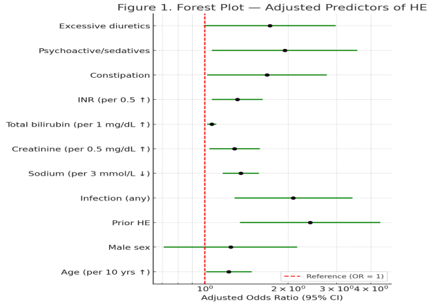
Table 3. Multivariable logistic regression of independent predictors of hepatic encephalopathy
| Predictor | Adjusted OR (95 % CI) | p-value |
|---|---|---|
| Age | 1.22 (1.01–1.48) | 0.040 |
| Male sex | 1.24 (0.71–2.16) | 0.44 |
| Prior HE | 2.41 (1.34–4.33) | 0.003 |
| Infection | 2.09 (1.28–3.43) | 0.003 |
| Sodium | 1.35 (1.16–1.57) | <0.001 |
| Creatinine | 1.28 (1.04–1.58) | 0.020 |
| Total bilirubin | 1.06 (1.02–1.10) | 0.010 |
| INR | 1.31 (1.06–1.62) | 0.012 |
| Constipation | 1.68 (1.02–2.77) | 0.041 |
| Psychoactive/sedatives | 1.95 (1.06–3.57) | 0.031 |
| Excessive diuretics | 1.72 (0.99–2.98) | 0.054 |
Model performance: AUC 0.83 (95 % CI 0.78–0.88), Brier score 0.17, calibration slope 0.96, intercept –0.03, Hosmer–Lemeshow p = 0.41.
A Bedside nomogram that converts these predictors into a personalized chance of HE within 48 hours as shown in Figure 2a–b:
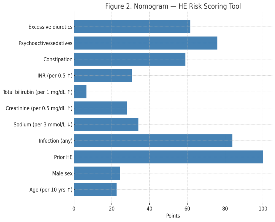
Figure 2 (a) Nomogram showing point allocation for each predictor
Figure 2 (b) Nomogram showing point allocation for each predicto
Figure 2 a & b. Nomogram illustrating significant predictors of hepatic encephalopathy for bedside risk estimation. The total score corresponds to the predicted probability of HE within 48 hours of hospital admission, allowing individualized clinical risk assessment.
DISCUSSION
In this prospective study conducted at a tertiary-care center in Mardan, Pakistan, 39.5% of hospitalized patients with cirrhosis developed hepatic encephalopathy (HE) either at presentation or within the first 48 hours of admission. International findings that suggest a 30–50% prevalence of HE among hospitalized cirrhotic patients are in close agreement with this frequency [13,14]. In line with extensive epidemiologic studies that demonstrate that early, lower-grade HE accounts for the majority of HE admissions and a substantial amount of the illness burden, the majority of cases in our cohort were categorized as Grade I–II [14,15].
With 36.6% and 29.7% of patients affected, respectively, infection and hyponatremia were the most frequent precipitants. These results are consistent with other cohorts in which infection accounts for 30–40% of HE admissions [18], but hyponatremia occurs in about 25–35% of patients and has been strongly linked to worse outcomes and increased risk [22]. Since illness and hyponatremia are both easily recognized and possibly curable precipitants, this pattern is especially pertinent in environments with limited resources. In contrast to many Western nations where MASLD currently predominates, Pakistan has a dual etiology of liver disease, with both hepatitis C virus (HCV) infection and MASLD significantly contributing to the advanced disease burden [6–10]. This helps to explain the high incidence of HE seen in our cohort.
Prior HE, active infection, lesser sodium, higher creatinine, higher bilirubin, higher INR, constipation, and exposure to psychoactive or sedative substances were all found to be independent predictors of HE by the multivariable model. These factors are indicative of well-established HE pathogenic processes. Previous research has clearly demonstrated that infection exacerbates ammonia neurotoxicity, increases blood–brain barrier permeability, and amplifies systemic inflammation [14–16,18]. The influence of hyponatremia on HE risk is well-established due to astrocyte swelling and osmotic dysregulation; the study's adjusted odds ratio of 1.35 per 3 mmol/L drop in sodium is in line with that of other international cohorts [15–17,22]. Numerous mechanistic and clinical studies have identified renal impairment, which is reflected in high creatinine, as a risk factor for HE. It contributes through decreased ammonia clearance and inflammatory amplification [17,18]. According to earlier prognostic research, elevated bilirubin and INR are indicative of declining synthetic function and are associated with HE risk as well as unfavorable outcomes in cirrhosis [19–21,24,25]. The probability of early HE was almost quadrupled by a history of previous HE, which supported relapse-prone patterns found in large multicenter trials like REACH [26]. The significance of treating these modifiable bedside factors, as advised by international recommendations, is highlighted by the fact that constipation and sedative exposure remained significant predictors [15,19,20].
Comparable to or better than previously published HE prediction models, the final model showed strong calibration and outstanding discrimination (AUC 0.83) [19,22,23]. Interestingly, the model's variables are all easily accessible at the time of hospitalization, which makes it an excellent tool for environments with minimal resources. The study gives clinicians a useful tool for early risk classification and triage by converting these indicators into a bedside nomogram. This strategy is consistent with organized HE care package studies that show that early, protocolized management improves outcomes [15, 23].
When compared to environments where MASLD is more prevalent, Pakistan's high HE burden is probably caused by the cohabitation of HCV and MASLD. Early detection of high-risk patients is essential in these settings, because there is a limited capacity for intensive care. Clear, doable targets for intervention are provided by infection, hyponatremia, renal impairment, and other recognized predictors. Incorporating this predictive model into ward-based care may facilitate early therapeutic interventions and more effective monitoring resource allocation.
This study has a number of advantages. It used standardized West Haven grading [2,13], was prospectively planned, included prespecified modeling with bootstrap validation, used systematic precipitant evaluation, and openly published both calibration and discrimination metrics. Beyond p-values, standardized mean differences were also employed to more accurately describe baseline disparities. The single-center design, absence of secret HE testing, and possible residual confounding are among the limitations. To evaluate generalizability, external validation from several centers is required. Future research should ascertain whether the nomogram can prevent ICU transfers, disease progression, and readmissions by initiating early HE care bundles, which include infection screening, salt correction, renal protection, laxative titration, and sedative evaluation [15,23].
CONCLUSION
Early HE in cirrhosis is frequent, predictable, and mostly caused by precipitants that can be changed. The variables found in this study offer significant locally relevant data from a high-burden LMIC scenario, while also closely matching worldwide findings. By using these variables to create an internally validated bedside nomogram, clinical outcomes and resource utilization can be maximized through early triage and focused care.
Author Contributions:
FUS, SA, RAK, RUD, NA:
Each author made substantial contributions to the conception and design of the study, or acquisition, analysis, and interpretation of data.
All authors were involved in drafting the manuscript or critically revising it for important intellectual content.
All authors approved the final version of the manuscript to be published and agree to be accountable for all aspects of the work.
REFERENCES
- Häussinger D, Dhiman RK, Felipo V, Görg B, Jalan R, Kircheis G, et al. Hepatic encephalopathy. Nat Rev Dis Primers. 2022;8(1):43. https://doi.org/10.1038/s41572-022-00366-6
- Vilstrup H, Amodio P, Bajaj J, Cordoba J, Ferenci P, Mullen KD, et al. Hepatic encephalopathy in chronic liver disease: 2014 Practice Guideline by AASLD and EASL. J Hepatol. 2014;61(3):642-659. https://doi.org/10.1016/j.jhep.2014.05.042
- Vilstrup H, Jepsen P, Andersen PK, Kamath PS. Hepatic encephalopathy and mortality in patients with cirrhosis. J Hepatol. 2020;73(4):1016-1022. https://doi.org/10.1016/j.jhep.2020.04.003
- Frenette CT, Mahmud N, Al-Kawas FH, Brenner K, Rich NE, Singal AG, et al. Hepatic encephalopathy-related hospitalizations in the United States: patient characteristics and outcomes. Dig Dis Sci. 2022;67(9):4393-4405. https://doi.org/10.1007/s10620-021-07075-2
- Kibble H, Elliott J, Coleman S, Hudson M, Neal TJ, Ruhashya C, et al. The assessment and management of cirrhotic patients with altered consciousness. United European Gastroenterol J. 2024;12(7):705-721. https://doi.org/10.1002/ueg2.12530
- Devarbhavi H, Asrani SK, Arab JP, Nartey YA, Pose E, Kamath PS. Global burden of liver disease: 2023 update. J Hepatol. 2023;79(2):516-537. https://doi.org/10.1016/j.jhep.2023.03.017
- Sepanlou SG, Safiri S, Bisignano C, Ikuta KS, Merat S, Saberifiroozi M, et al. The global, regional, and national burden of cirrhosis by cause in 195 countries and territories, 1990-2017. Lancet Gastroenterol Hepatol. 2020;5(3):245-266. https://doi.org/10.1016/S2468-1253(19)30349-8
- Dopico E, Khan A, Penon JL, Broll S, McElligott G, Rahman MM, et al. Prevalence of hepatitis C virus infection, genotypes and associated risk factors in Pakistan: a systematic review and meta-analysis. Infect Drug Resist. 2022;15:4465-4488. https://doi.org/10.2147/IDR.S367715
- Hassan F, Hashmi I, Ahmed W, Wahab S, Raza SA, Masood A, et al. Prevalence of non-alcoholic fatty liver disease in Pakistan: a systematic review and meta-analysis. Sci Rep. 2024;14:16156. https://doi.org/10.1038/s41598-024-70481-9
- Younossi ZM, Golabi P, de Avila L, Paik JM, Srishord M, Fukui N, et al. The global epidemiology of NAFLD and NASH in patients with type 2 diabetes: a systematic review and meta-analysis. J Hepatol. 2019;71(4):793-801. https://doi.org/10.1016/j.jhep.2019.06.021
- Bellafante D, De Sibio MT, Xiao J, Trawiński K, Nardelli S, Hidalgo R, et al. The management of hepatic encephalopathy from ward to intensive care unit. J Clin Med. 2023;13(1):166. https://doi.org/10.3390/jcm13010166
- Lauridsen MM, Bajaj JS. Inpatient management of hepatic encephalopathy. Clin Liver Dis (Hoboken). 2024;23(1):e0105. https://doi.org/10.1097/CLD.0000000000000105
- Ferenci P, Lockwood A, Mullen K, Tarter R, Weissenborn K, Blei AT. Hepatic encephalopathy—definition, nomenclature, diagnosis, and quantification: final report of the working party at the 11th World Congress of Gastroenterology, Vienna, 1998. Hepatology. 2002;35(3):716-721. https://doi.org/10.1053/jhep.2002.31250
- Butterworth RF. Pathogenesis of hepatic encephalopathy and brain edema in acute liver failure. J Clin Exp Hepatol. 2015;5(Suppl 1):S96-S103. https://doi.org/10.1016/j.jceh.2014.02.004
- European Association for the Study of the Liver. EASL Clinical Practice Guidelines on the management of hepatic encephalopathy. J Hepatol. 2022;77(3):807-824. https://doi.org/10.1016/j.jhep.2022.06.001
- Bass NM, Mullen KD, Sanyal A, Poordad F, Neff G, Leevy CB, et al. Rifaximin treatment in hepatic encephalopathy. N Engl J Med. 2010;362(12):1071-1081. https://doi.org/10.1056/NEJMoa0907893
- Wong F. Acute kidney injury in liver cirrhosis: new definition and application. Nat Rev Nephrol. 2021;17(2):137-138. https://doi.org/10.1038/s41581-020-00370-0
- Piano S, Brocca A, Mareso S, Angeli P. Infections complicating cirrhosis. Liver Int. 2018;38(Suppl 1):126-133. https://doi.org/10.1111/liv.13649
- Nabi E, Bajaj JS. Useful tests for hepatic encephalopathy in clinical practice. Curr Gastroenterol Rep. 2020;22(9):43. https://doi.org/10.1007/s11894-020-00780-7
- Tapper EB, Parikh ND. Mortality due to cirrhosis and liver cancer in the United States, 1999-2016: observational study. BMJ. 2018;362:k2817. https://doi.org/10.1136/bmj.k2817
- Ahluwalia V, Wade JB, Thacker LR, White MB, Gavis EA, Fagan A, et al. Differential impact of hyponatremia and hepatic encephalopathy on health-related quality of life and brain metabolite abnormalities in cirrhosis. J Hepatol. 2018;69(2):310-318. https://doi.org/10.1016/j.jhep.2018.02.026
- Nardelli S, Lattanzi B, Torrisi S, Greco F, Gioia S, Merli M. Clinical outcomes of a structured HE bundle in hospitalized cirrhotic patients. Liver Int. 2022;42(2):356-364. https://doi.org/10.1111/liv.15142
- Cordoba J, Ventura-Cots M, Simón-Talero M, Amoros A, Pavesi M, Vilstrup H, et al. Characteristics, risk factors, and mortality of cirrhotic patients hospitalized for hepatic encephalopathy with and without acute-on-chronic liver failure (ACLF). J Hepatol. 2014;60(2):275-281. https://doi.org/10.1016/j.jhep.2013.09.010