
Biocompatibility and Cytotoxicity of Universal
Dental Adhesives on Human Dental Pulp Cells:
A Systematic Review of in Vitro Studies
Correspondence and article notes 8
ABSTRACT
Background: Unpolymerized monomers released from resin-based universal dental adhesives may diffuse through dentinal tubules towards the pulp, and cell-culture studies suggest that some monomers can impair pulp-cell function. Previous systematic reviews have addressed resin composites or dental adhesives in general, often with mixed cell types; to our knowledge none has restricted its question to human dental pulp cells exposed to universal adhesives. How the in vitro effects of universal adhesives on human pulp cells compare between products and experimental conditions therefore remains unclear.
Objective: To systematically review in vitro studies reporting the effects of universal dental adhesives on human dental pulp cells, compared with untreated controls or other adhesives, on cell viability/metabolic activity, cell death and inflammatory mediators, and to describe how these outcomes varied with the experimental conditions reported.
Methods: PubMed, ScienceDirect and Google Scholar were last searched on for in vitro studies published between 1 January 2020 and 23rd April 2026. English-language, full-text studies using human dental pulp cells and universal adhesives were eligible. Two reviewers independently screened studies and extracted data. Methodological quality and completeness of reporting were appraised with an author-developed six-domain checklist adapted from CONSORT items (not a validated risk-of-bias tool). We synthesized findings narratively; we did not conduct a meta-analysis or formal certainty-of-evidence grading. The review was reported according to PRISMA 2020. This review was not registered in PROSPERO or any other registry
Results: Seven in vitro studies were included (five used human dental pulp stem cells; two used human pulp cells). Viability or metabolic activity was assessed by MTT (five studies), WST-1 (one), trypan blue (one) and Live/Dead staining (one); flow cytometry was used in two. Exposure ranged from 24 hours to 7 days, and three studies assessed more than one time point. Three studies measured inflammatory mediators (ELISA, MAGPIX or qPCR). Results differed markedly between products and between studies. At 24 hours, metabolic activity was close to control for Single Bond Universal and Clearfil Universal Bond Quick but markedly reduced for OptiBond Universal, ZipBond Universal, Bond Force II and Futurabond DC. Cytokine changes were reported after ZipBond Universal (IL-1β, IL-6, TNF-α and IL-10 increased) and Single Bond Universal (IL-8 increased). Findings for isolated monomers depended on concentration and could not be generalized to commercial adhesives.
Conclusion: Across seven heterogeneous in vitro studies, universal adhesives affected human pulp cells differently by product, concentration, assay, and exposure time. These data do not identify compositional determinants of biocompatibility, do not allow products to be ranked, and are not evidence of clinical pulpal safety or harm. Standardized protocols and clinical studies are needed.
Keywords: Biocompatible Material; Cytotoxicity Tests; Dental Bonding; Dental Pulp Cells
INTRODUCTION
Biocompatibility is commonly defined as the ability of a material to perform its intended function in a specific application with an appropriate host response [1,2]. For resin-based dental adhesives applied to dentin, relevant biological responses include cell viability and metabolic activity, genotoxicity, and inflammatory and repair-related responses of the dentin–pulp complex [2,3]. Preserving pulp vitality is a cornerstone of restorative dentistry, as the dental pulp provides immune surveillance, dentinogenic and sensory functions [3]. Resin-based adhesive restorations rely on bonding systems that underpin minimal-intervention dentistry [4]. A biological concern with these restorations is that unpolymerized monomers may diffuse through dentinal tubules, particularly in deep cavity preparations, and reach pulp cells [5,6]. Diffusion through residual dentin differs from direct exposure after pulp exposure: in the first, dentin thickness, tubule patency, and dentinal fluid flow attenuate the amount of monomer reaching the pulp, whereas in the second, the adhesive resin contacts pulp tissue directly [6]. When residual dentin is thin, this barrier is reduced, which may increase the potential for pulpal exposure to monomers at concentrations that are harmful to cells in experimental models [4-6]. These are potential biological implications, not established clinical consequences of universal adhesive exposure.
Because of the interest in clinical simplification, universal adhesives have become widely used in clinical practice, as they can be applied in etch-and-rinse, self-etch or selective enamel-etch mode without changing the product formula [7,8]. These systems combine priming and bonding in a single bottle, reducing steps, technique sensitivity, and chair-side time [7,8]. Their formulations, however, usually contain several methacrylate monomers, including 2-hydroxyethyl methacrylate (HEMA), triethylene glycol dimethacrylate (TEGDMA), bisphenol A glycidyl methacrylate (Bis-GMA) and urethane dimethacrylate (UDMA), and functional acidic monomers such as 10-methacryloyloxydecyl dihydrogen phosphate (10-MDP) [9,10], together with relatively high solvent concentrations that can impair polymerization efficiency and increase water sorption [9,10,11].
In vitro studies attribute methacrylate-based adhesive cytotoxicity largely to unreacted monomers remaining when polymerization is incomplete [2]. Residual HEMA, TEGDMA and Bis-GMA can elute from resin-based materials [10,11] and, in experimental models, may reach sufficient concentrations to impair cell function [1] Mechanisms reported in cell-culture studies include mitochondrial dysfunction with ATP depletion, increased production of reactive oxygen species (ROS) with glutathione depletion, and activation of apoptotic pathways [12]. These mechanisms are specific to the monomer, concentration and cell type studied. For example, TEGDMA was reported to induce both caspase-dependent and caspase-independent apoptosis in pulp cells [13], whereas the evidence for HEMA and for part of the TEGDMA evidence comes from macrophages [14], and cannot be assumed to apply to pulp cells.
Monomer exposure has also been associated, in cell-culture studies, with altered production of cytokines such as interleukin-1β (IL-1β), interleukin-6 (IL-6) and tumor necrosis factor-α (TNF-α) in pulpal cell [15,16] and with changes in matrix metalloproteinases (MMP-2, MMP-8), their inhibitor TIMP-1 and the odontoblast-associated protein DMP-1[16]. These are cellular and molecular findings. These studies have not established whether they translate into pulpitis or impaired reparative dentine formation in vivo.
Clinical outcomes are a separate question. Clinical trials have evaluated postoperative sensitivity after composite restorations placed with different adhesive strategies [17, 18, 19]; these studies assess clinical symptoms rather than cellular mechanisms and were not designed to determine whether monomer cytotoxicity causes them. Cell-culture findings can therefore suggest, but cannot confirm, a biological basis for clinical pulpal responses.
Previous systematic reviews differ in scope from the present question. Wiertelak-Makała et al. [20] reviewed 14 in vitro studies (2017–2023) of methacrylate-based resin composites using human and murine cell lines, so their review addressed restorative composites rather than adhesives and did not restrict the cell type to the pulp. Other reviews discussed how methodological variation (extract preparation, cell type, assay conditions, and exposure time) complicates comparisons of adhesive cytotoxicity but did not synthesize evidence from human pulp cells for universal adhesives. The unresolved question is therefore how universal adhesives compare in their reported effects on human dental pulp cells, and how consistent those effects are across experimental conditions. This review addresses that question narrowly and treats the variation between studies as a finding in itself.
Research question and objective: In in vitro studies using human dental pulp cells (population), what effects are reported after exposure to universal dental adhesives or their extracts (intervention), compared with untreated controls or other adhesives (comparator), on cell viability/metabolic activity, cell death, inflammatory mediators and related outcomes (outcomes), and how consistent are these effects across products and experimental conditions?
METHODS
Protocol and registration. This systematic review was conducted and reported in accordance with the PRISMA 2020 statement; the completed checklist is provided as a supplementary file. The authors prepared a protocol specifying the research question, search strategy, eligibility criteria, and data-extraction methods before screening. The protocol was not registered in PROSPERO or any other registry. We do not regard registration as necessary for in vitro reviews; the absence of prospective registration is acknowledged as a limitation because readers cannot verify that methods and outcomes were pre-specified.
The inclusion criteria were: (1) in vitro studies assessing the cytotoxicity or biocompatibility of universal dental adhesives; (2) use of human dental pulp cells (primary cultures or established cell lines); (3) use of a recognized cytotoxicity or viability assay (for example MTT, Alamar Blue, WST-1, trypan blue, LDH release, Live/Dead staining or flow cytometry); (4) explicit reporting of exposure conditions, concentrations and quantitative results; (5) publication or online-ahead-of-print availability between 1 January 2020 and 30 December 2025 (the Epub date was used where it preceded issue assignment); and (6) availability of the full text in English. The exclusion criteria were: (1) in vivo or animal studies; (2) in vitro studies using non-pulpal cell types (for example, gingival fibroblasts, osteoblast-like cells, macrophages or epithelial cells) or non-human cells; (3) review articles, case reports and conference abstracts; (4) studies lacking quantitative cytotoxicity data; (5) experimental adhesive compositions in which the cytotoxicity of the base adhesive could not be assessed independently; and (6) studies that did not specify the adhesive brand or composition. We applied English-language and full-text restrictions for feasibility (reviewers' language proficiency, no translation resources) and because full text was needed to extract exposure conditions and quantitative data; this may introduce language and availability bias.
Information sources and search strategy: We searched PubMed (MEDLINE; pubmed.ncbi.nlm.nih.gov), ScienceDirect (Elsevier), and Google Scholar.
Other databases (Scopus, Web of Science, Embase) were considered but not searched; this may have led to missed studies. PubMed: ("universal adhesive*"[tiab] OR "universal dental adhesive*"[tiab] OR "universal bonding agent*"[tiab] OR "multi-mode adhesive*"[tiab] OR "multimode adhesive*"[tiab] OR "Dentin-Bonding Agents"[MeSH Terms]) AND ("cytotoxic*"[tiab] OR "biocompatib*"[tiab] OR "cell viability"[tiab] OR "MTT"[tiab] OR "Cell Survival"[MeSH Terms] OR "Biocompatible Materials"[MeSH Terms]) AND ("Dental Pulp"[MeSH Terms] OR "dental pulp cell*"[tiab] OR "pulp stem cell*"[tiab] OR "DPSC*"[tiab] OR "Odontoblasts"[MeSH Terms]); filters: publication date 2020/01/01 to 2026/04/23; language English. ScienceDirect: ("universal adhesive" OR "universal adhesives" OR "multi-mode adhesive") AND ("dental pulp" OR "pulp cells" OR "pulp stem cells") AND (cytotoxicity OR "cell viability" OR biocompatibility); limits: years 2020–2026, article type research articles (language screened manually). Google Scholar: "universal adhesive" "dental pulp" cytotoxicity viability; custom range 2020–2026.
Study selection: We exported records to Mendeley, identified duplicates, and removed them. Two reviewers independently screened all records by title and abstract and then independently assessed potentially eligible full texts against the inclusion and exclusion criteria. We resolved disagreements through discussion and, if consensus was not reached, by consulting a third reviewer. The numbers of records screened, full texts assessed and studies included, with reasons for full-text exclusion, are shown in the PRISMA 2020 flow diagram (Figure 1).
Extracted items were: study characteristics (authors, year, country); adhesive brand, type, manufacturer and composition; cell source, passage number and culture conditions; exposure model (adhesive extract, monomer solution or modified formulation), dilution/concentration and exposure time; assay type; quantitative viability values and IC50 where reported; inflammatory/apoptotic outcome data; polymerization conditions (light-curing unit, irradiation time); and statistical comparisons. Missing or unclear data were recorded as not reported (NR) and were not imputed. Where values were available only graphically, approximate values were read from the figures and marked with †.
Outcomes. Primary outcomes were (a) cell viability/metabolic activity and (b) cell death/cytotoxicity, which were extracted and reported separately because they are related but not interchangeable. For (a), MTT, WST-1 and Alamar Blue assays measure mitochondrial/metabolic activity as a proxy for viable cell number, and trypan blue exclusion measures membrane integrity; values were extracted as percentage of control. A reduction in metabolic activity does not by itself establish cell death. For (b), we measured direct indicators: Live/Dead staining, propidium iodide (PI)-based flow cytometry (apoptosis/necrosis), and LDH release. Secondary outcomes were (c) inflammatory mediators (IL-1/IL-1β, IL-6, IL-8, IL-10 and TNF-α, measured as protein by ELISA or multiplex immunoassay [MAGPIX], or as mRNA by qPCR, and extracted as concentration or fold-change relative to control); (d) matrix-remodeling and odontoblast-associated markers (TIMP-1, MMPs, DMP-1), which were extracted separately and not classed as inflammatory mediators; and (e) factors that may influence biocompatibility, namely degree of conversion, monomer release and oxidative stress, extracted descriptively where reported.
Methodological quality and risk of bias. No single validated risk-of-bias tool is universally accepted for in vitro dental-materials studies. We did not apply the QUIN tool, used in a previous review of resin composites [2]. Two reviewers independently appraised each study using an author-developed checklist adapted from selected CONSORT items; disagreements were resolved by discussion or a third reviewer. CONSORT is a reporting guideline, not a risk-of-bias instrument, and its adaptation here has not been validated. We therefore interpreted the checklist as an appraisal of methodological quality and completeness of reporting; these relate to, but are distinct from, risk of bias (the likelihood of systematic error in a study's results), and the scores must not be read as a risk-of-bias judgement. The six domains, each chosen because it bears on interpretation or reproducibility of in vitro cytotoxicity data, were: (1) origin and characterization of pulp cells (0–2; reporting/external validity); (2) justification of sample size (0–1; precision); (3) dose–response analysis (0–2; design); (4) replication of key experiments (0–2; random error); (5) appropriate statistical analysis (0–2; analysis); and (6) reporting of polymerization conditions (0–2; standardization of exposure). The maximum score was 11. Author-defined categories were: higher quality/more complete reporting, 9–11 points; moderate, 5–8; lower, ≤4. Scores were not used to exclude studies or to weight the synthesis.
Synthesis methods: Studies were grouped (i) by outcome (viability/metabolic activity; cell death; inflammatory and matrix-related markers), (ii) within each outcome by adhesive product and exposure time, and (iii) by exposure model (adhesive extract, monomer solution or modified formulation). We present results study by study in structured tables, and we did not perform vote counting or effect-size conversion. A meta-analysis was not undertaken because the outcomes were not sufficiently comparable: results were reported in different metrics (percentage of control, fold-change, relative units, or not reported); assays measured different constructs; extract preparation, dilution or concentration, curing and exposure times differed or were not reported; cell sources differed; few products were tested under common conditions in more than one study; and variability and sample sizes were often not reported, so a pooled estimate would not have been interpretable. Reporting bias was not formally assessed (see Results).
RESULTS
The searches identified 347 records (PubMed 124, ScienceDirect 156, Google Scholar 67). After removing 89 duplicate records, we screened 258 records by title and abstract and excluded 215. The full texts of 43 reports were assessed. (Figure 1).
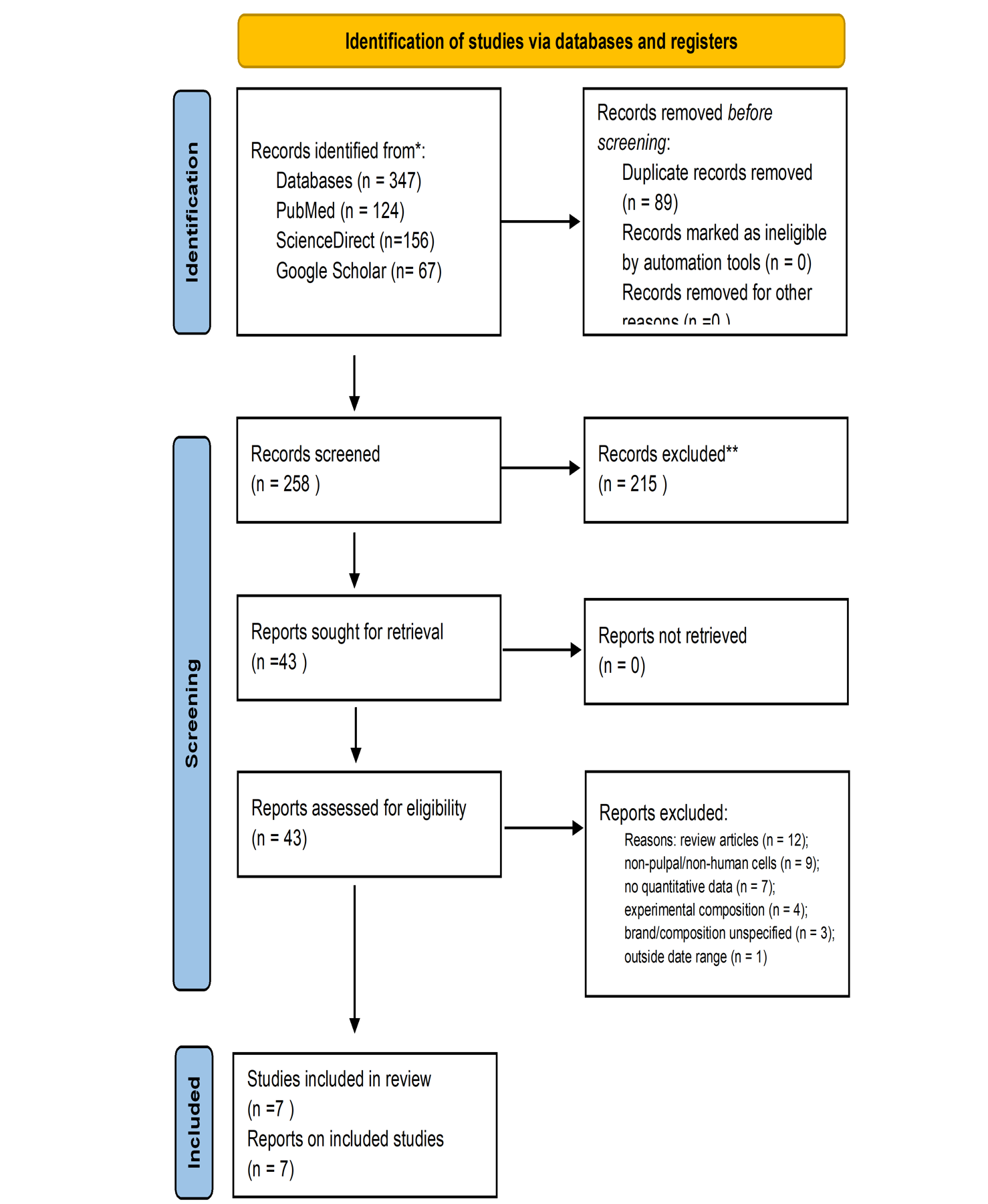
Text in the figure
Identification of studies via databases and registers IdentificationRecords removed before screeningDuplicate records n = 89)0 )Records removed before screeningRecords marked as ineligible by automation tools (n = 0)Databases (n = 347)Google Scholar (n= 67)PubMed (n = 124) Records screened(n = Reports sought for retrieval(n =43 )Reports not retrieved(n = 0) Reports assessed for eligibility(n = 43)
Table 1 highlight study characteristics and outcomes included studies. Values marked † were read approximately from figures. The quality score in the last column (out of 11) derives from checklist presented in Table 3.
Table 1: Summary of Studies on Dental Adhesives and Cytotoxicity
Study |
Adhesives / Materials Evaluated | Cell Type | Assays Used | Exposure Duration |
Concentration / Dilution | Cell Viability (% of control, mean ± SD/SE or range) | IC₅₀ | Apoptosis / Cytokine Values (mean ± SD/SE or fold-change vs control) | p-values (vs control) | Quality (Table 3) | |
|---|---|---|---|---|---|---|---|---|---|---|---|
| Ersöz et al. [16] | BFII, CS3UB, G2BU, PBU, OBU, GBU | Human dental pulp cells | MTT, ELISA | 24–72 h | 1:1, 1:2, 1:5 dilutions in culture medium | 72 h (1:1): BFII 83.5 ± 14.9; CS3UB 54.7 ± 3.4; G2BU 51.7 ± 5.8; PBU 73.8 ± 4.3; OBU 64.2 ± 7.1; GBU 95.8 ± 11.6. 48 h (1:1): BFII 91.4 ± 9.1; CS3UB 76.4 ± 9.2; G2BU 71.9 ± 11.3; PBU 77.9 ± 5.6; OBU 80.2 ± 8.2; GBU 96.8 ± 11.1. 24 h (1:1): BFII 101.1 ± 17.1; CS3UB 93.6 ± 9.1; G2BU 85.2 ± 8.2; PBU 88.6 ± 8.6; OBU 89.2 ± 7.8; GBU 107.8 ± 12.5 |
NR | TNF-α: increased at 24 h, decreased at 72 h (p<0.05); no difference between adhesives (p>0.05). IL-1: increased at 24 h and 72 h (p<0.05). TIMP-1: decreased at 24 h and 72 h (p<0.05). DMP-1: increased at 24 h (p<0.05), NS at 72 h. Exact pg/mL NR. | p<0.05 for time effect; p>0.05 for between-adhesive differences in TNF-α | 11/11 | |
| Lima et al. [15] | SBU, OBU, ZBU | hDPSCs | MTT, MAGPIX | 24 h | Extract of cured adhesive in medium (exact dilution NR) | 24 h (MTT, % of control): Control 100; SBU ~100† (NS vs control); OBU ~28†; ZBU ~28†. Both OBU and ZBU drastically reduced mitochondrial metabolism vs control and SBU. | NR | IL-1β: Control ~0.7, SBU ~0.9, OBU ~0.8, ZBU ~2.2; IL-6: Control ~2.2, SBU ~2.5, OBU ~1.8, ZBU ~5.0; IL-8: Control ~25, SBU ~100, OBU ~15, ZBU ~20; TNF-α: Control ~1.2, SBU ~1.5, OBU ~1.2, ZBU ~2.8; IL-10: Control ~1.5, SBU ~1.8, OBU ~1.6, ZBU ~7.0. ZBU significantly increased IL-1β, IL-6, TNF-α, IL-10 vs control (p<0.05). SBU significantly increased IL-8 vs control (p<0.05). | p<0.05 for OBU/ZBU MTT vs control; p<0.05 for ZBU cytokines vs control | 10/11 | |
| de Lucena et al. [21] | TiO₂ nanotube-modified adhesives (CHX-loaded, DOX-loaded, blank) | hDPSCs | MTT, Live/Dead | 24 h | 5 wt% TiO₂-NT in universal adhesive | No significant cytotoxicity observed vs control for any formulation. Exact % NR. | NR | Live/Dead: no significant increase in dead cells vs control. Exact counts NR. | p>0.05 (NS vs control) | 10/11 | |
| Monteiro et al. [22] | G-IEMA vs Bis-GMA adhesives | Human pulp cells | MTT, flow cytometry (PI) | 24 h | Monomer solutions 1–4 mM; polymerized & non-polymerized adhesive extracts | MTT: G-IEMA viability was +20% higher than Bis-GMA in non-polymerized extracts and +51% higher in polymerized extracts. Absolute % NR. | NR | Flow cytometry: G-IEMA was less cytotoxic than Bis-GMA. Apoptosis/necrosis (PI): lower cell death with G-IEMA vs Bis-GMA. Exact % NR. | p<0.05 (G-IEMA vs Bis-GMA) | 9/11 | |
| Koruyucu et al. [23] | Futurabond M, Clearfil S3, G Premio Bond | hDPSCs | Trypan blue | 24–72 h | Extract of cured adhesive in medium (exact dilution NR) | 24 h: MTA (pos. control) 87.2%; G Premio Bond 57%; Clearfil S3 59.5%; Futurabond M 83%. 48 h & 72 h: no significant differences between groups. | NR | NR | p<0.05 at 24 h (G Premio, Clearfil S3 vs MTA/control); p>0.05 at 48 h and 72 h | 11/11 | |
| Kazak et al. [24] | CUBQ, BFII, FBDC | hDPSCs | WST-1 | 24 h – 7 days | Extract of cured adhesive in medium (exact dilution NR) | 24 h: Control 100.00 ± 0.00; CUBQ 98.47 ± 8.93; BFII 20.51 ± 2.89; FBDC 7.20 ± 0.34. 72 h: Control 100.00 ± 0.00; CUBQ 94.25 ± 10.00; BFII 14.02 ± 0.58; FBDC 16.12 ± 1.05. 7 d: Control 100.00 ± 0.00; CUBQ 22.39 ± 3.80; BFII 17.16 ± 3.48; FBDC 18.35 ± 3.61. |
NR | NR | p<0.05 for BFII and FBDC vs control at all time points; p>0.05 for CUBQ at 24 h and 72 h | 10/11 | |
| Cavallaro-Mota et al. [25] | 10-MDP vs GPDM monomers | hDPSCs | MTT, qPCR, flow cytometry | 24 h | 10-MDP: 1–3 mM; GPDM: 1–4 mM in DMSO (≤0.1%) | MTT (% of control): 10-MDP at 3 mM: 35.0 (65% reduction, p<0.05). GPDM at 1–4 mM: ~80.0 (20% reduction, p<0.05 at 1.25 mM). Flow cytometry: 10-MDP at 3 mM: ~12.0% cell death (6% increase over control ~6.0%). GPDM at 4 mM: ~10.5% cell death. | 10-MDP: 3 mM | 10-MDP: reduced IL-6 and IL-8 (more pronounced at 3 mM, p<0.05); no change in IL-1β, IL-10, TNF-α. GPDM: increased IL-1β (all conc., p<0.05); decreased IL-10 (all conc., p<0.05); reduced IL-6 (at 1 and 3 mM, p<0.05); reduced IL-8 (at 2 and 3 mM, p<0.05); no effect on TNF-α. | p<0.05 for stated cytokine changes vs control | 10/11 | |
Abbreviations: hDPSCs, human dental pulp stem cells; NR, not reported; NS, not significant; †, approximate value read from a figure; IC₅₀, half-maximal inhibitory concentration; BFII, Bond Force II; CS3UB, Clearfil S3 Bond Universal; G2BU, G2-Bond Universal; PBU, Prime&Bond Universal; OBU, OptiBond Universal; GBU, Gluma Bond Universal; SBU, Single Bond Universal; ZBU, ZipBond Universal; CUBQ, Clearfil Universal Bond Quick; FBDC, Futurabond DC; MTA, mineral trioxide aggregate (positive control); G-IEMA, dendrimeric methacrylate monomer [define as in Monteiro et al.]; Bis-GMA, bisphenol A glycidyl methacrylate; 10-MDP, 10-methacryloyloxydecyl dihydrogen phosphate; GPDM, glycerol phosphate dimethacrylate; TiO₂-NT, titanium dioxide nanotubes; CHX, chlorhexidine; DOX, doxycycline; MTT, 3-(4,5-dimethylthiazol-2-yl)-2,5-diphenyltetrazolium bromide; WST-1, water-soluble tetrazolium salt-1; MAGPIX, multiplex bead immunoassay; PI, propidium iodide; ELISA, enzyme-linked immunosorbent assay; qPCR, quantitative polymerase chain reaction; DMP-1, dentin matrix protein-1; TIMP-1, tissue inhibitor of metalloproteinases-1. Quality category: author-developed checklist (Table 3); not a validated risk-of-bias judgement.
Table 2: Study-specific summary of reported viability/metabolic-activity and inflammatory findings by adhesive product (not a ranking)
| Adhesive | Key monomers (descriptive) | Viability / metabolic activity (study-specific) | Inflammatory / cell-death findings | Study context [ref] |
|---|---|---|---|---|
| Gluma Bond Universal (GBU) | MDP, HEMA | 95.8% of control at 72 h (1:1), 96.8% at 48 h, 107.8% at 24 h; highest of the six adhesives tested in this study [16] | Across all adhesives: TNF-α and IL-1 changed over time, no between-adhesive difference in TNF-α; TIMP-1 decreased [16] | Human dental pulp cells; MTT, ELISA; 24–72 h; 1:1–1:5 dilutions [16] |
| Single Bond Universal (SBU) | MDP, HEMA, dimethacrylates | About 100% of control at 24 h, not significantly different [15] | IL-8 significantly increased vs control [15] | hDPSCs; MTT, MAGPIX; 24 h; extract, dilution NR [15] |
| Futurabond M | HEMA, acidic monomers, UDMA | 83% at 24 h (positive control MTA 87.2%); no significant differences between groups at 48 and 72 h [23] | Not assessed (trypan blue only) [23] | hDPSCs; trypan blue; 24–72 h; extract, dilution NR [23] |
| Futurabond DC (FBDC) | 7.20 ± 0.34% (24 h), 16.12 ± 1.05% (72 h), 18.35 ± 3.61% (7 d); p<0.05 vs control at all time points [24] | NR [27] | hDPSCs; WST-1; 24 h–7 d; extract, dilution NR [24] | |
| Clearfil Universal Bond Quick (CUBQ) | MDP, hydrophilic amide monomer | 98.47 ± 8.93% (24 h), 94.25 ± 10.00% (72 h), 22.39 ± 3.80% (7 d); not significantly different from control at 24 and 72 h [24] | NR [27] | hDPSCs; WST-1; 24 h–7 d; extract, dilution NR [24] |
| Clearfil S3 Bond / Clearfil S3 Bond Universal (CS3UB) | 59.5% at 24 h, p<0.05 vs control/MTA, no significant differences at 48–72 h [23]; CS3UB 54.7 ± 3.4% at 72 h (1:1) [16] | Not assessed [23]; CS3UB: see GBU row for study-wide findings [16] | hDPSCs, trypan blue [23]; human pulp cells, MTT, ELISA [16]. [Verify whether the two products are identical] | |
| G Premio Bond | MDP, HEMA, dimethacrylate | 57% at 24 h, p<0.05 vs MTA/control; no significant differences at 48–72 h [23]. Not the lowest viability reported across studies (see Futurabond DC and Bond Force II) | Not assessed [23] | hDPSCs; trypan blue; 24–72 h; extract, dilution NR [23] |
| Bond Force II (BFII) | HEMA, Bis-GMA | 83.5 ± 14.9% at 72 h (1:1) [16]; 20.51 ± 2.89% (24 h), 14.02 ± 0.58% (72 h), 17.16 ± 3.48% (7 d), p<0.05 [24]. Results differ markedly between studies | Study-wide findings, see GBU row [16]; NR [24] | Human pulp cells, MTT, ELISA [16]; hDPSCs, WST-1 [24] |
| OptiBond Universal (OBU) | GPDM, HEMA, Bis-GMA | About 28% of control at 24 h (MTT), p<0.05 [15]; 64.2 ± 7.1% at 72 h (1:1) [16]. Reduced metabolic activity; cell death not directly assessed | No significant cytokine increase vs control reported [15]; study-wide findings, see GBU row [16] | hDPSCs; MTT, MAGPIX [4]; human pulp cells; MTT, ELISA [16] |
| Prime&Bond Universal (PBU) | PENTA, HEMA | 73.8 ± 4.3% at 72 h (1:1) [16] | Study-wide findings, see GBU row [16] | Human pulp cells; MTT, ELISA; 24–72 h [16] |
| G2-Bond Universal (G2BU) | 51.7 ± 5.8% at 72 h (1:1); 71.9 ± 11.3% at 48 h; 85.2 ± 8.2% at 24 h [16] [verify product name] | Study-wide findings, see GBU row [16] | Human pulp cells; MTT, ELISA; 24–72 h [16] | |
| ZipBond Universal (ZBU) | HEMA, MDP, Bis-GMA | About 28% of control at 24 h (MTT), p<0.05 [15] | IL-1β, IL-6, TNF-α and IL-10 significantly increased vs control [15] | hDPSCs; MTT, MAGPIX; 24 h; extract, dilution NR [15] |
Abbreviations as in Table 1. Values are as reported in the cited studies and are study-specific (different cells, assays, extract preparation, concentrations and times); they must not be compared across studies or read as a ranking. Monomer information is descriptive.
Table 3: Methodological quality and completeness-of-reporting appraisal of included studies (author-developed checklist; see note)
| Study | Cell Origin/ Characteristics (0–2) |
Sample-Size Justification (0–1) |
Dose–Response Analysis (0–2) |
Replication of Experiments (0–2) |
Statistical Evaluation (0–2) |
Polymerization Conditions (0–2) |
Total score (max 11) | Quality category |
|---|---|---|---|---|---|---|---|---|
| Ersöz et al. [16] | 2 | 1 | 2 | 2 | 2 | 2 | 11 | Higher |
| Lima et al. [15] | 2 | 1 | 1 | 2 | 2 | 2 | 10 | Higher |
| de Lucena et al. [21] | 2 | 1 | 2 | 1 | 2 | 2 | 10 | Higher |
| Monteiro et al. [22] | 2 | 1 | 1 | 2 | 2 | 1 | 9 | Higher |
| Koruyucu et al. [23] | 2 | 1 | 2 | 2 | 2 | 2 | 11 | Higher |
| Kazak et al. [24] | 2 | 1 | 1 | 2 | 2 | 2 | 10 | Higher |
| Cavallaro-Mota et al. [25] | 2 | 0 | 2 | 2 | 2 | 2 | 10 | Higher |
Author-developed checklist adapted from selected CONSORT items; it appraises methodological quality and completeness of reporting and has not been validated as a risk-of-bias tool. Maximum score 11. Categories: higher quality/more complete reporting 9–11; moderate 5–8; lower ≤4.
Methodological quality and completeness of reporting (by domain).
Table 3 shows by domain: origin and characterization of cells was fully reported in all seven studies (2/2) and statistical analysis was fully scored in all seven (2/2); sample-size justification was reported in six studies (score 1) and not in Cavallaro-Mota et al. [25] (score 0); dose–response analysis scored 2 in four studies [16,21,23,25] and 1 in three [15,22,24]; replication scored 2 in six studies and 1 in de Lucena et al. [21]; and polymerization conditions scored 2 in six studies and 1 in Monteiro et al. [22]. These scores reflect reporting and design features and should not be interpreted as low risk of bias; several studies did not report extract dilution, variability or exact values (Table 1).
The included studies did not test dentin barrier thickness or degree of conversion; none tested adhesive diffusion through dentin or compared different degrees of polymerization
Synthesis of results by outcome. Viability/metabolic activity. Results varied widely between products and studies (Tables 1 and 2). In Ersöz et al. [16], viability at 72 hours (1:1 dilution) ranged from 51.7% (G2-Bond Universal) and 54.7% (Clearfil S3 Bond Universal) to 95.8% (Gluma Bond Universal). In Lima et al. [15], MTT activity at 24 hours was about 100% of control for Single Bond Universal but about 28% for OptiBond Universal and ZipBond Universal (p <0.05). In Koruyucu et al. [23], viability at 24 hours was 83% for Futurabond M, 59.5% for Clearfil S3 and 57% for G Premio Bond (the latter two, p <0.05), with no significant differences at 48 and 72 hours. In Kazak et al. [24], Clearfil Universal Bond Quick was close to control at 24 and 72 hours (98.5% and 94.3%) but fell to 22.4% at 7 days, whereas Bond Force II (20.5%, 14.0%, 17.2%) and Futurabond DC (7.2%, 16.1%, 18.4%) were markedly reduced at all time points (p<0.05). Modified adhesives containing TiO₂ nanotubes showed no significant cytotoxicity at 24 hours [21]. The same product could give very different results in different studies: Bond Force II 83.5% at 72 hours [16] versus 14.0% at 72 hours [21]; OptiBond Universal 64.2% at 72 hours [16] versus about 28% at 24 hours [15].
Cell death. Only three studies measured cell death directly. Live/Dead staining showed no significant increase in dead cells after TiO₂-nanotube-modified adhesives [21]. In flow-cytometry studies, a G-IEMA-based formulation produced less cell death than a Bis-GMA-based formulation [22]; in Cavallaro-Mota et al. [25], 10-MDP at 3 mM was associated with about 12% cell death (control about 6%) and GPDM at 4 mM with about 10.5%. In the latter study, MTT activity fell to about 35% of control with 10-MDP at 3 mM but was about 80% with GPDM at 1–4 mM, so the monomers did not show a uniform pattern, and reduced metabolic activity exceeded measured cell death. Findings for isolated monomers depend on concentration and cannot be attributed to commercial adhesives containing several monomers.
Inflammatory mediators and matrix markers. In Ersöz et al. [16], TNF-α increased at 24 hours and decreased at 72 hours, and IL-1 increased at both times (p<0.05), with no difference between adhesives in TNF-α; TIMP-1 decreased at 24 and 72 hours, and DMP-1 increased at 24 hours with no significant change at 72 hours (exact concentrations not reported). In Lima et al. [15], ZipBond Universal significantly increased IL-1β, IL-6, TNF-α and IL-10 and Single Bond Universal significantly increased IL-8 relative to control (approximate values, graph-read). In Cavallaro-Mota et al. [25], 10-MDP reduced IL-6 and IL-8 without changing IL-1β, IL-10 or TNF-α, whereas GPDM increased IL-1β, decreased IL-10 and reduced IL-6 and IL-8 at some concentrations without affecting TNF-α. TIMP-1 was measured in one study only [16].
Time depends on the subject. Results at different time points were not consistent: group differences at 24 hours disappeared by 48–72 hours in Koruyucu et al. [23]; Clearfil Universal Bond Quick showed a delayed reduction at 7 days; and viability and cytokine changes in Ersöz et al. [16] varied with time. Four studies assessed only 24 hours [15,21,22,25], so we could not examine these patterns.
Reporting bias. Reporting bias was not formally assessed. We did not use funnel plots or Egger's test because they require an adequate number of studies and comparable effect estimates, and with seven heterogeneous in vitro studies and no meta-analysis, they would have limited utility. Therefore, selective reporting and publication bias (for example, under-reporting of non-significant cytotoxicity results) cannot be excluded, and the extracted data may be affected, as several values were not reported.
DISCUSSION
This systematic review synthesized seven in vitro studies of the effects of universal adhesives, and in two studies of isolated monomers or experimental formulations, on human dental pulp cells. The principal finding is variability: reported viability and cytokine responses differed between products, concentrations, assays, and exposure times, and the same product sometimes gave very different results across studies. The extracted data support the following study-specific observations: metabolic activity at 24 hours close to control for Single Bond Universal [15] and Clearfil Universal Bond Quick [24] but markedly reduced for OptiBond Universal and ZipBond Universal [15] and for Bond Force II and Futurabond DC; and cytokine changes after ZipBond Universal and Single Bond Universal [15]. Separate in vitro investigations have shown that certain Universal adhesive formulations induce genotoxic and cytotoxic effects, although some used non pulpal cellular models [26,27].
Contradictory results and methodological heterogeneity. Several results conflict. Bond Force II retained 83.5% viability at 72 hours in one study [16] but 14.0% in another [24], and OptiBond Universal gave 64.2% at 72 hours [16] versus about 28% at 24 hours [15]. Differences in methodology, a recognized problem in biocompatibility testing [26,27], may contribute: (i) cell type and source (hDPSCs versus primary pulp cells) can differ in metabolic activity and stress resistance; (ii) adhesive presentation (neat extracts, serial dilutions, monomer solutions or nanotube-modified formulations) alters the concentration delivered to cells, and extract dilution was not reported in several studies; (iii) light-curing units, irradiation times and serum presence were not uniform and can influence degree of conversion and monomer release; and (iv) assay type and exposure time differ. Because most products were tested in only one study, these factors cannot be separated, and none of them was shown to explain the discrepancies.
Viability, cell death and inflammatory response are distinct outcomes. In Cavallaro-Mota et al. [25], 10-MDP at 3 mM reduced MTT activity to about 35% of control while flow cytometry showed about 12% cell death (control about 6%). Inflammatory outcomes also did not simply follow viability: in Lima et al. [15], Single Bond Universal did not reduce MTT activity but increased IL-8, whereas ZipBond Universal both reduced MTT activity and increased IL-1β, IL-6, TNF-α and IL-10. Viability assays alone may therefore not capture inflammatory responses. Unpolymerized resin monomers can cause oxidative stress and mitochondrial dysfunction in pulpal cells [28].
Monomers, composition and mechanisms. Two included studies examined monomers or experimental monomer-based formulations [22,25]. In Cavallaro-Mota et al. [25], 10-MDP at 3 mM reduced viability more than GPDM at 1–4 mM, yet the cytokine profiles differed (GPDM increased IL-1β and decreased IL-10, whereas 10-MDP reduced IL-6 and IL-8). In Monteiro et al. [22], a G-IEMA-based formulation showed higher viability than a Bis-GMA-based one. Mechanistic literature outside the included studies reports that unpolymerized methacrylate monomers can cause oxidative stress, mitochondrial dysfunction, and apoptosis in cultured cells; much of this evidence comes from non-pulpal cells, and the included studies did not measure these mechanisms [28-31].
Matrix-related markers and time dependence. Only one included study [16] measured TIMP-1 and DMP-1: TIMP-1 decreased at 24 and 72 hours, and DMP-1 increased at 24 hours with no significant change at 72 hours, across the adhesives tested. Time-dependent patterns also differed between studies (differences at 24 hours that disappeared by 48–72 hours [23]; a delayed reduction at 7 days for Clearfil Universal Bond Quick [24]), and four studies assessed 24 hours only. Single-time-point designs may therefore miss early or delayed effects.
Dentin barrier and clinical relevance. The studies included applied extracts or monomers directly to cultured cells and therefore model direct contact; none examined dentin thickness or dentinal fluid. Residual dentin attenuates, but may not abolish, monomer passage: an in vitro dentin-slice study reported that bonding-agent components diffusing through dentin slices still produced ROS and apoptosis in pulpal cells [32]. No included study showed that universal adhesives cause pulpitis or postoperative sensitivity, and clinical trials of postoperative sensitivity address different outcomes [17,33].
Limitations: The evidence base comprises seven in vitro studies, with most products being tested in a single study. Two studies tested isolated monomers or experimental formulations rather than commercial products, and some included products may not be universal adhesives under every classification.
Limitations of the review process. The literature search covered studies published between January 2020 and April 2026; therefore, relevant studies published subsequently were not included. The exact final search date should be specified for transparency and reproducibility.
Implications for research. Future studies should use standardized extract preparation (for example, according to ISO 10993-5) and report dilutions, curing conditions, sample sizes and variability; combine a metabolic assay with a direct measure of cell death; include several time points; compare commercial products under identical conditions; and use dentin-barrier models. Clinical studies are needed to determine whether any in vitro differences are relevant to pulpal outcomes.
CONCLUSION
Within the limits of seven heterogeneous in vitro studies, the effects of universal dental adhesives on human dental pulp cells differed between products, concentrations, assays and exposure times. The data do not allow product grading or conclusions about clinical pulpal safety
Author Contributions:
HHS, MAG, KAM, MSS, GH, KS
Each author made substantial contributions to the conception and design of the study, or acquisition, analysis, and interpretation of data.
All authors were involved in drafting the manuscript or critically revising it for important intellect content.
All authors approved the final version of the manuscript to be published and agree to be accountable for all aspects of the work.
REFERENCES
- Moharamzadeh K, Brook IM, Van Noort R. Biocompatibility of resin-based dental materials. Materials (Basel). 2009;2(2):514-48. https://doi.org/10.3390/ma2020514.
- Franca CM, de Souza Balbinot G, Cunha D, Saboia VD, Ferracane J, Bertassoni LE. In-vitro models of biocompatibility testing for restorative dental materials: from 2D cultures to organs on-a-chip. Acta Biomater. 2022;150:58-66. https://doi.org/10.1016/j.actbio.2022.07.060.
- Brizuela M, Bordoni B. Anatomy, head and neck, dental pulp. In: StatPearls [Internet]. Treasure Island (FL): StatPearls Publishing; 2026 Jan–. Updated 2025 Dec 9. Available from: https://www.ncbi.nlm.nih.gov/books/NBK537112/.
- Caldas IP, Alves GG, Barbosa IB, Scelza P, de Noronha F, Scelza MZ. In vitro cytotoxicity of dental adhesives: a systematic review. Dent Mater. 2019;35(2):195-205. https://doi.org/10.1016/j.dental.2018.11.028.
- Hamid A, Hume WR. The effect of dentine thickness on diffusion of resin monomers in vitro. J Oral Rehabil. 1997;24(1):20-5. https://doi.org/10.1046/j.1365-2842.1997.00490.x.
- Bouillaguet S. Biological risks of resin-based materials to the dentin-pulp complex. Crit Rev Oral Biol Med. 2004;15(1):47-60. https://doi.org/10.1177/154411130401500105.
- Bourgi R, Kharouf N, Cuevas-Suárez CE, Lukomska-Szymanska M, Haikel Y, Hardan L. A literature review of adhesive systems in dentistry: key components and their clinical applications. Appl Sci. 2024;14(18):8111. https://doi.org/10.3390/app14188111.
- Breschi L, Maravic T, Mazzitelli C, Josic U, Mancuso E, Cadenaro M, et al. The evolution of adhesive dentistry: from etch-and-rinse to universal bonding systems. Dent Mater. 2025;41(2):141-58. https://doi.org/10.1016/j.dental.2024.11.011.
- Ahmed MH, Yoshihara K, Nagaoka N, Yao C, Matsukawa A, Yoshida Y, et al. Acrylamide monomers in universal adhesives. Dent Mater. 2023;39(3):246-59. https://doi.org/10.1016/j.dental.2023.01.003.
- Delgado AH, Ahmed MH, Ferreira MN, Azul AM, Polido M, Yoshihara K, et al. Physico-chemical properties and performance of functional monomers used in contemporary dental adhesive technology. J Adhes Dent. 2025;27:175-93. https://doi.org/10.3290/j.jad.c_2297.
- Almusa A, Delgado AH, Ashley P, Young AM. Determination of dental adhesive composition throughout solvent drying and polymerization using ATR-FTIR spectroscopy. Polymers (Basel). 2021;13(22):3886. https://doi.org/10.3390/polym13223886.
- Xu S, Chen L, Lin X, Yang X, He L, Yan S, et al. Resin monomers induce apoptosis of the pulp-dentin complex through the mitochondrial pathway. J Toxicol Sci. 2024;49(12):531-41. https://doi.org/10.2131/jts.49.531.
- Lovász BV, Berta G, Lempel E, Sétáló G Jr, Vecsernyés M, Szalma J. TEGDMA (triethylene glycol dimethacrylate) induces both caspase-dependent and caspase-independent apoptotic pathways in pulp cells. Polymers (Basel). 2021;13(5):699. https://doi.org/10.3390/polym13050699.
- Krifka S, Hiller KA, Spagnuolo G, Jewett A, Schmalz G, Schweikl H. The influence of glutathione on redox regulation by antioxidant proteins and apoptosis in macrophages exposed to 2-hydroxyethyl methacrylate (HEMA). Biomaterials. 2012;33(21):5177-86. https://doi.org/10.1016/j.biomaterials.2012.04.013.
- Lima AF, Oliveira AA, Fronza BM, Braga RR, Andia DC. Toxicity and cytokine release from human dental pulp stem cells after exposure to universal dental adhesives cured by single peak and polywave LEDs. Dent Mater. 2024;40(5):837-41. https://doi.org/10.1016/j.dental.2024.03.004.
- Ersöz B, Aydin N, Oktay EA, Çal İK, Karaoğlanoğlu S. Effects of universal adhesives on dentin matrix proteins, matrix metalloproteinases and cytokine release of human pulp cells. Odontology. 2026;114(1):138-48. https://doi.org/10.1007/s10266-025-01107-3.
- Javed K, Noor N, Nasir MZ, Manzoor MA. Comparison of postoperative hypersensitivity between total-etch and universal adhesive system: a randomized clinical trial. Sci Rep. 2024;14(1):678. https://doi.org/10.1038/s41598-024-51175-8.
- Fang K, Chen K, Shi M, Wang L. Effect of different adhesive systems on dental defects and sensitivity to teeth in composite resin restoration: a systematic review and meta-analysis. Clin Oral Investig. 2023;27(6):2495-511. https://doi.org/10.1007/s00784-023-05007-0.
- Polesso Patias M, Fernandes-e-Silva P, Carreño NL, Lund RG, Piva E, da Silva AF, et al. Comparative clinical performance of universal adhesives versus etch-and-rinse and self-etch adhesives: a meta-analysis. Clin Oral Investig. 2025;29(7):352. https://doi.org/10.1007/s00784-025-06427-w.
- Wiertelak-Makała K, Szymczak-Pajor I, Bociong K, Śliwińska A. Considerations about cytotoxicity of resin-based composite dental materials: a systematic review. Int J Mol Sci. 2024;25(1):152. https://doi.org/10.3390/ijms25010152.
- de Lucena FS, Bronze-Uhle ÉS, Logan M, Lewis S, de Oliveira Gallinari M, Lisboa Filho PN, et al. Modification of a universal adhesive with CHX- or DOX-loaded TiO₂ nanotubes as an anti-MMP strategy. Dent Mater. 2026;42(3):460-72. https://doi.org/10.1016/j.dental.2025.11.006.
- Monteiro D, Barbier M, Delgado AH, Gonçalves L, Chaves-Ferreira M, e Cruz JV, et al. A multifunctional dendrimer for BPA-free dental adhesives: polymerization behavior and cytotoxic profile of G-IEMA. Int J Adhes Adhes. 2025;143:104162. https://doi.org/10.1016/j.ijadhadh.2025.104162.
- Koruyucu M, Akay C, Solakoglu S, Gencay K. Investigation of the cytotoxic effect of current dentine bonding agents on human dental pulp cells. BMC Oral Health. 2024;24(1):1207. https://doi.org/10.1186/s12903-024-04985-1.
- Kazak M, Sarialioglu Gungor A, Ozman Z, Donmez N. Comparative cell viability of dentin-bonding adhesive systems on human dental pulp stem cells: time-dependent analysis. BMC Oral Health. 2024;24(1):663. https://doi.org/10.1186/s12903-024-04438-9.
- Cavallaro-Mota FD, Esposo GN, Kury M, Fronza BM, Saraceni CH, Andia DC, et al. Assessment of 10-MDP and GPDM monomers on viability and inflammatory response in human dental pulp stem cells. Dent Mater. 2025;41(1):1-6. https://doi.org/10.1016/j.dental.2024.10.008.
- Wawrzynkiewicz A, Rozpedek-Kaminska W, Galita G, Lukomska-Szymanska M, Lapinska B, Sokolowski J, et al. The cytotoxicity and genotoxicity of three dental universal adhesives: an in vitro study. Int J Mol Sci. 2020;21(11):3950. https://doi.org/10.3390/ijms21113950.
- Sürmelioğlu D, Hepokur C, Yavuz SA, Aydın U. Evaluation of the cytotoxic and genotoxic effects of different universal adhesive systems. J Conserv Dent. 2020;23(4):384-9. https://doi.org/10.4103/JCD.JCD_376_20.
- Jiao Y, Ma S, Wang Y, Li J, Shan L, Liu Q, et al. N-acetyl cysteine depletes reactive oxygen species and prevents dental monomer-induced intrinsic mitochondrial apoptosis in vitro in human dental pulp cells. PLoS One. 2016;11(1):e0147858. https://doi.org/10.1371/journal.pone.0147858.
- Jiao Y, Niu T, Liu H, Tay FR, Chen JH. Protection against HEMA-induced mitochondrial injury in vitro by Nrf2 activation. Oxid Med Cell Longev. 2019;2019:3501059. https://doi.org/10.1155/2019/3501059.
- Nocca G, Callà C, Martorana GE, Cicillini L, Rengo S, Lupi A, et al. Effects of dental methacrylates on oxygen consumption and redox status of human pulp cells. Biomed Res Int. 2014;2014:956579. https://doi.org/10.1155/2014/956579.
- Baldion PA, Velandia-Romero ML, Castellanos JE. Dental resin monomers induce early and potent oxidative damage on human odontoblast-like cells. Chem Biol Interact. 2021;333:109336. https://doi.org/10.1016/j.cbi.2020.109336.
- Lee BS, Jan YD, Huang GS, Huang CH, Chou HY, Wang JS, et al. Effect of dentin bonding agent diffusing through dentin slices on the reactive oxygen species production and apoptosis of pulpal cells. J Formos Med Assoc. 2015;114(4):339-46. https://doi.org/10.1016/j.jfma.2012.12.009.
- Castro AS, Maran BM, Gutierrez MF, Chemin K, Mendez-Bauer ML, Bermúdez JP, et al. Effect of dentin moisture in posterior restorations performed with universal adhesive: a randomized clinical trial. Oper Dent. 2022;47(2):E91-105. https://doi.org/10.2341/20-215-C.